. 2019 Oct 8;29(2):301-316.e10.
doi: 10.1016/j.celrep.2019.08.104.

Tau-Mediated Disruption of the Spliceosome Triggers Cryptic RNA Splicing and Neurodegeneration in Alzheimer's Disease

Affiliations

Tau-Mediated Disruption of the Spliceosome Triggers Cryptic RNA Splicing and Neurodegeneration in Alzheimer's Disease

Yi-Chen Hsieh et al. Cell Rep. .

Abstract

In Alzheimer's disease (AD), spliceosomal proteins with critical roles in RNA processing aberrantly aggregate and mislocalize to Tau neurofibrillary tangles. We test the hypothesis that Tau-spliceosome interactions disrupt pre-mRNA splicing in AD. In human postmortem brain with AD pathology, Tau coimmunoprecipitates with spliceosomal components. In Drosophila, pan-neuronal Tau expression triggers reductions in multiple core and U1-specific spliceosomal proteins, and genetic disruption of these factors, including SmB, U1-70K, and U1A, enhances Tau-mediated neurodegeneration. We further show that loss of function in SmB, encoding a core spliceosomal protein, causes decreased survival, progressive locomotor impairment, and neuronal loss, independent of Tau toxicity. Lastly, RNA sequencing reveals a similar profile of mRNA splicing errors in SmB mutant and Tau transgenic flies, including intron retention and non-annotated cryptic splice junctions. In human brains, we confirm cryptic splicing errors in association with neurofibrillary tangle burden. Our results implicate spliceosome disruption and the resulting transcriptome perturbation in Tau-mediated neurodegeneration in AD.

Keywords: Alzheimer’s disease; RNA splicing; SmB; Tau; U1-70K; cryptic splicing; intron retention; neurodegeneration; neurofibrillary tangles; spliceosome.


Conflict of interest statement

DECLARATION OF INTERESTS

The authors declare no competing interests.

Figures

Figure 1.
Figure 1.. Tau Associates and Genetically Interacts with Spliceosomal Factors
(A) Following coimmunoprecipitation, 1,065 Tau-associated proteins were identified from AD or control postmortem brain tissue homogenates (n = 4 each). Volcano plot highlights proteins showing >1.5-fold increased (red) or decreased (green) interactions with Tau in AD (all p < 0.05), including numerous ribonucleoproteins (p = 7.7 × 10⁻⁵). Spliceosome components are denoted. See also Figure S1, Tables S1, and Data S1, tabs i and ii. (B) Compared with controls (Ctrl, top left), TauV337M expression in the eye (GMR-GAL4) causes reduced eye size and roughened surface (bottom left: 1.98 ± 0.08, mean score ± SEM, n > 35 for all genotypes), and RNAi targeting spliceosome factors enhanced toxicity (bottom row, p < 0.0001: SmB, 3.67 ± 0.06; SmD2, 4.00 ± 0.00; U1–70K, 4.04 ± 0.03; U1C, 3.92 ± 0.09; and SmE, 4.00 ± 0.00). RNAi lines were non-toxic when expressed independently (top row). See also Data S1, tab iii. (C) TauWT expression in the adult retina (Rh1-GAL4) causes reduced electroretinogram (ERG) amplitude in 5-day-old flies, and this phenotype is enhanced in SmBMG heterozygotes (n > 10 for quantification). See also Figures S2A–S2C. (D) Pan-neuronal expression of TauR406W (elav-GAL4) causes progressive neuropil vacuolization (arrows) in hematoxylin and eosin-stained frontal brain sections in 10-day-old adult flies, and this phenotype is enhanced in SmBMG heterozygotes (n > 8 for quantification). Scale bar: 50 μm. See also Figures S2D and S2E. All error bars denote mean ± SEM. **p < 0.01; ***p < 0.001; ns, not significant.
Figure 2.
Figure 2.. Disruption of the Spliceosome following Tau Expression in Drosophila
(A and B) Pan-neuronal human TauR406W disrupts expression of multiple spliceosomal proteins in 1-day-old adult fly head homogenates. Western blots (A) were probed for Tau or spliceosome proteins and normalized to the loading control, GAPDH (n = 4 replicates for quantification, B). The Y12 antibody recognizes both SmB and SmD3. (C) mRNA expression was also examined in 1-day-old adult heads (n = 3). (D and E) Whole-mount stains of 1-day-old adult fly brains (D) reveal depletion of SmB/D3 and U1A/SNF protein (red, n = 15 for quantification, E). Nuclei are colabeled with 4′,6-diamidino-2-phenylindole (DAPI; grayscale). Scale bar: 20 μm. (F) Glial expression of TauWT (repo-GAL4) induces cytoplasmic foci (arrowheads) of SmB/D3 (Y12, red) that colocalize with phospho-Tau aggregates (green) in 10-day-old adult brains. Nuclei are labeled with DAPI (blue). Boxed region is magnified at right. Quantification (n > 9) reveals 14.86% ± 2.3% of phospho-Tau aggregates colabeling for Sm proteins; aggregates were not observed in controls. Scale bar: 10 μm. See also Figure S3A. All error bars denote mean ± SEM. *p < 0.05; **p < 0.01; ***p < 0.001; ns, not significant.
Figure 3.
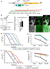
Figure 3.. Loss of SmB, Encoding a Core Spliceosome Factor, Causes Reduced Survival and Progressive Locomotor Impairment
(A) The SmB locus, including single coding exon (CDS, yellow), untranslated regions (UTRs, gray), and transposable elements. To generate SmBMG, recombination-mediated cassette exchange was performed using SmBMI, introducing a coding exon for GFP, flanked by flexible linkers (Ls) and splice acceptor and donor sequences (SA/SDs). The deficiency strain (red), Df(2L)BSC453, deletes ~51 kb including the entire SmB locus. The transgenic genomic rescue strain, SmBGR, carries a ~90-kb bacterial artificial chromosome (BAC, CH321–75P02, green), including SmB. See also Table S2. (B) SmBMG/MG homozygotes demonstrate expression of the GFP-SmB fusion protein at levels comparable with controls (n = 3 for quantification, SmB protein normalized to Actin). (C) GFP-SmB (green) is localized to the nucleus (DAPI, grayscale) in brains from SmBMG/MG adults. Scale bar: 20 μm. Boxed region (top) is magnified below. (D) SmBMG/MG adults (black) exhibit reduced survival and this phenotype is partially rescued by the SmB genomic rescue (blue) (n > 313 per genotype). See also Figure S4A. (E) SmBMG/MG adults also manifest progressive locomotor impairment (n > 5 groups). See also Figure S4B. (F and G) Both the SmBMG/MG survival (F, n > 288) and locomotor (G, n > 4 groups) phenotypes were rescued by pan-neuronal expression of wild-type SmB. All error bars denote mean ± SEM. *p < 0.05; **p < 0.01; ***p < 0.001; ns, not significant.
Figure 4.
Figure 4.. Loss of SmB Causes Neurodegeneration
(A) Hematoxylin and eosin-stained frontal brain section at the level of mushroom body calyx, highlighting the region of interest for (B) and (C). Scale bar: 25 μm. (B and C) SmBMG/MG adults (B) show progressive loss of cortical nuclei between 2 and 25 days of age (n > 5 for quantification, C). Scale bar: 25 μm. (D) Frontal section at the level of lamina highlighting region of interest for (E) and (F) and DAB-positive cholinergic neurons (ChAT>lacZ reporter). Scale bar: 20 μm. (E and F) SmBMG/MG animals (E) exhibit cholinergic neuron loss between 2 and 25 days (n > 9 for quantification, F). Scale bar: 10 μm. All error bars denote mean ± SEM. *p < 0.05; **p < 0.01; ***p < 0.001; ns, not significant. See also Figures S4C–S4F and S5.
Figure 5.
Figure 5.. Tau Expression and SmB Loss Cause Similar Splicing Errors in Drosophila Brains
(A) Key for splicing errors shown in (B). (B) Pan-neuronal expression of TauWT or TauR406W induces differential expression of cryptic splicing errors (left) and intron retention (right), similar to SmBMG/MG, based on RNA-seq in 1-, 10-, or 20-day-old flies (n = 3 per genotype, except n = 2 for same-batch control for TauR406W comparison). See also Figure S6B, Tables S4 and S5, and Data S1, tabs iv and v. (C) Histograms for representative Tau-induced splicing errors in 20-day-old flies, showing RNA-seq reads normalized between control and TauR406W and split reads spanning splice junctions. Transcript structures are indicated below, including reference-annotated (black) versus aberrant exons (gray) resulting from cryptic splice junctions. Transcript orientation (arrows) and the 5′-splice donor sites (green bars) analyzed in (F) are denoted. See also Figure S6C. (D) Genes harboring splicing errors following pan-neuronal expression of TauR406W (union of 1-, 10-, and 20-day results) strongly overlap with SmBMG/MG (10 days). Percentage denotes Tau-associated, differentially spliced genes that also overlap. See also Figures S6D–S6F. (E) Splicing errors in TauR406W transgenic and SmBMG/MG flies occur more commonly in genes with greater numbers of introns (left) or annotated alternatively spliced transcripts (right), compared with all Drosophila genes. ***p < 0.001. (F) Splicing errors occur at exon-intron junctions with splice donor sequences that diverge from the consensus motif for U1 spliceosome binding. The consensus splice donor (5′-splice site) sequence (bold) is shown, along with splice donor sequences corresponding to splicing errors in representative genes (green bars in C). Nucleotides that diverge from the consensus are shown (red), along with the splice site binding strength score. See also Figures S7A and S7B. (G) Many splicing errors in TauR406W flies affect protein coding sequences (blue) or untranslated regions (UTRs: green, 5′-UTR; yellow: 3′-UTR), and are likely to disrupt protein expression. See also Data S1, tabs vi and vii.
Figure 6.
Figure 6.. Hypothetical Model for Tau-Spliceosome Interactions in AD
We propose that spliceosome factors can associate with either insoluble or soluble Tau species, respectively, leading to cytoplasmic sequestration or disrupting snRNP assembly and/or stability. Tau-spliceosome interactions likely contribute to splicing errors, global transcriptome perturbation and, ultimately, CNS dysfunction and neuronal loss.

References

    1. Abreha MH, Dammer EB, Ping L, Zhang T, Duong DM, Gearing M, Lah JJ, Levey AI, and Seyfried NT (2018). Quantitative Analysis of the Brain Ubiquitylome in Alzheimer’s Disease. Proteomics 18, e1800108. - PMC - PubMed
    1. Ali YO, Ruan K, and Zhai RG (2012). NMNAT suppresses tau-induced neurodegeneration by promoting clearance of hyperphosphorylated tau oligomers in a Drosophila model of tauopathy. Hum. Mol. Genet 21, 237–250. - PMC - PubMed
    1. Ambadipudi S, Biernat J, Riedel D, Mandelkow E, and Zweckstetter M (2017). Liquid-liquid phase separation of the microtubule-binding repeats of the Alzheimer-related protein Tau. Nat. Commun 8, 275. - PMC - PubMed
    1. Andrews S (2010). FastQC: a quality control tool for high throughput sequence data.http://www.bioinformatics.babraham.ac.uk/projects/fastqc.
    1. Anne J (2010). Arginine methylation of SmB is required for Drosophila germ cell development. Development 137, 2819–2828. - PubMed

Publication types

MeSH terms

LinkOut - more resources