

Investigation of In Vitro Antioxidant and Antibacterial Potential of Silver Nanoparticles Obtained by Biosynthesis Using Beech Bark Extract
- PMID: 31597312
- PMCID: PMC6827055
- DOI: 10.3390/antiox8100459
Investigation of In Vitro Antioxidant and Antibacterial Potential of Silver Nanoparticles Obtained by Biosynthesis Using Beech Bark Extract
Abstract
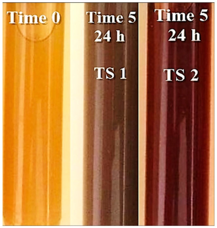
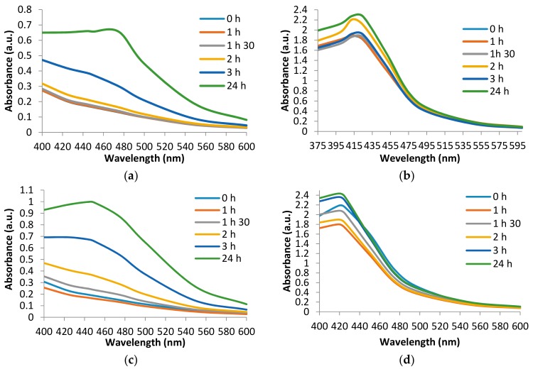
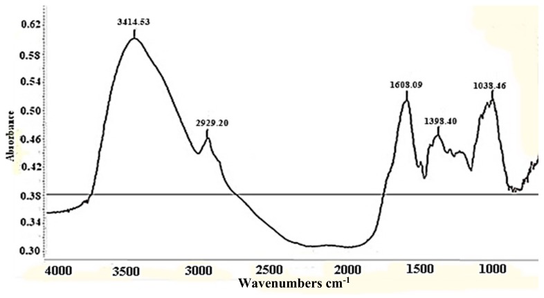
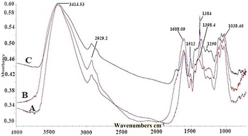
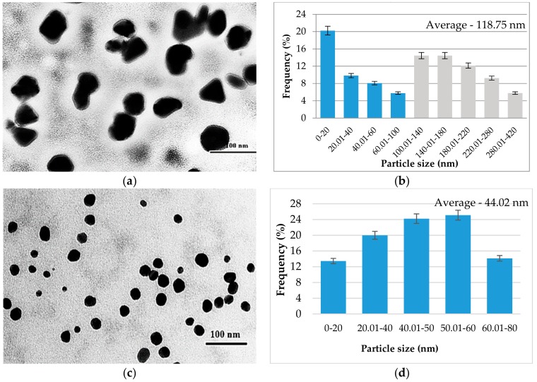
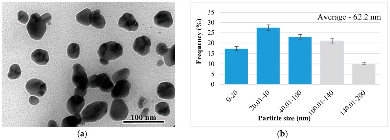
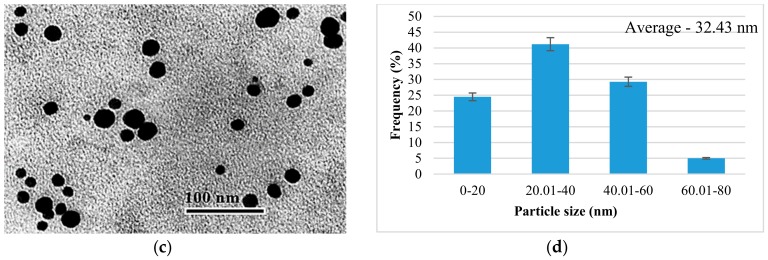
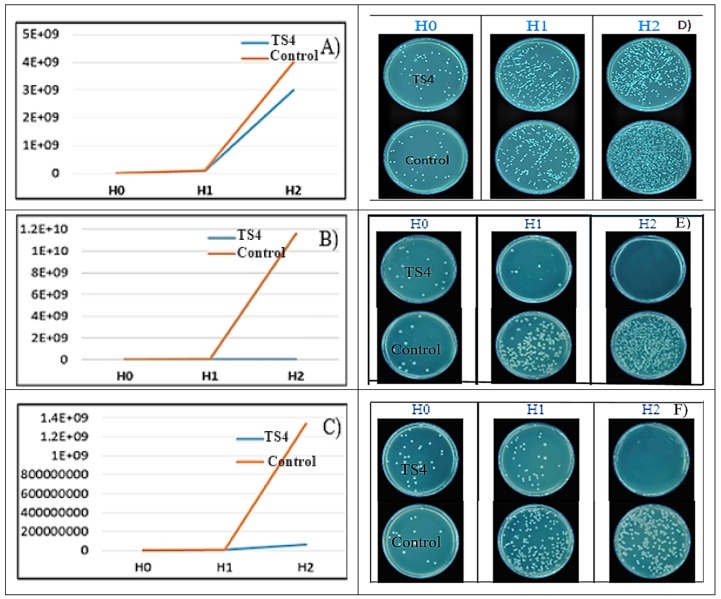
Green synthesis is one of the rapid and best ways for silver nanoparticles (AgNP) synthesis. In the present study, synthesis and bioactivity of AgNPs has been demonstrated using water beech (Fagus sylvatica L.) bark extract. The physical and chemical factors such as time, metal ion solution, and pH, which play a vital role in the AgNPs synthesis, were assessed. The AgNPs were characterized by ultraviolet-visible (UV-Vis) spectrometry, Fourier transform infrared spectroscopy (FT-IR), and transmission electron microscopy (TEM). Antioxidant and antimicrobial activity of the obtained AgNPs was evaluated. AgNPs were characterized by color change pattern, and the broad peak obtained at 420-475 nm with UV-Vis confirmed the synthesis of AgNPs. FT-IR results confirmed that phenols and proteins of beech bark extract are mainly responsible for capping and stabilization of synthesized AgNPs. TEM micrographs showed spherical or rarely polygonal and triangular particles with an average size of 32 nm at pH = 9, and 62 nm at pH = 4. Furthermore, synthesized AgNPs were found to exhibit antioxidant activity and have antibacterial effect against Staphylococcus aureus, methicillin-resistant Staphylococcus aureus (MRSA), Escherichia coli, and Pseudomonas aeruginosa. These results indicate that bark extract of F. sylvatica L. is suitable for synthesizing stable AgNPs, which act as an excellent antimicrobial agent.
Keywords: antibacterial; antioxidant; beech bark; polyphenols; silver nanoparticles.
Conflict of interest statement
The authors declare no conflict of interest.
Figures
References
-
- Chaturvedi V., Verma P. Fabrication of silver nanoparticles from leaf extract of Butea monosperma (flame of forest) and their inhibitory effect on bloom-forming cyanobacteria. Bioresour. Bioprocess. 2015;2:18. doi: 10.1186/s40643-015-0048-6. - DOI
-
- Kouvaris P., Delimitis A., Zaspalis V., Papadopoulos D., Tsipas S.A., Michailidis N. Green synthesis and characterization of silver nanoparticles produced using arbutus unedo leaf extract. Mater. Lett. 2012;76:18–20. doi: 10.1016/j.matlet.2012.02.025. - DOI
-
- Ahmad N., Sharma S. Green synthesis of silver nanoparticles using extracts of Ananas comosus. Green Sustain. Chem. 2012;2:141–147. doi: 10.4236/gsc.2012.24020. - DOI
-
- Prabhu S., Poulose E.K. Silver nanoparticles: Mechanism of antimicrobial action, synthesis, medical applications, and toxicity effects. Int. Nano Lett. 2012;2:32. doi: 10.1186/2228-5326-2-32. - DOI
-
- Parlinska-Wojtan M., Kus-Liskiewicz M., Depciuch J., Sadik O. Green synthesis and antibacterial effects of aqueous colloidal solutions of silver nanoparticles using camomile terpenoids as a combined reducing and capping agent. Bioprocess Biosyst. Eng. 2016;39:1213–1223. doi: 10.1007/s00449-016-1599-4. - DOI - PMC - PubMed
LinkOut - more resources
Full Text Sources
