. 2019 Oct 8;11(10):920.
doi: 10.3390/v11100920.

Internal Ribosome Entry Site Dramatically Reduces Transgene Expression in Hematopoietic Cells in a Position-Dependent Manner

Affiliations

Internal Ribosome Entry Site Dramatically Reduces Transgene Expression in Hematopoietic Cells in a Position-Dependent Manner

Qingyun Zheng et al. Viruses. .

Abstract

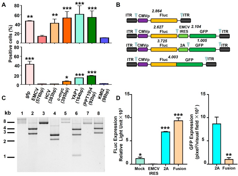
Bicistronic transgene expression mediated by internal ribosome entry site (IRES) elements has been widely used. It co-expresses heterologous transgene products from a message RNA driven by a single promoter. Hematologic gene delivery is a promising treatment for both inherited and acquired diseases. A combined strategy was recently documented for potential genome editing in hematopoietic cells. A transduction efficiency exceeding ~90% can be achieved by capsid-optimized recombinant adeno-associated virus serotype 6 (rAAV6) vectors. In this study, to deliver an encephalomyocarditis virus (EMCV) IRES-containing rAAV6 genome into hematopoietic cells, we observed that EMCV IRES almost completely shut down the transgene expression during the process of mRNA-protein transition. In addition, position-dependent behavior was observed, in which only the EMCV IRES element located between a promoter and the transgenes had an inhibitory effect. Although further studies are warranted to evaluate the involvement of cellular translation machinery, our results propose the use of specific IRES elements or an alternative strategy, such as the 2A system, to achieve bicistronic transgene expression in hematopoietic cells.

Keywords: bicistronic transgene; gene therapy; hematopoietic cells; internal ribosome entry site (IRES); recombinant adeno-associated virus vector.


Conflict of interest statement

The authors declare no conflict of interest.

Figures

Figure 1
Figure 1
Capsid-optimized recombinant adeno-associated virus serotype 6 (rAAV6) vectors represented the most efficient gene delivery method for hematopoietic cells. (A) K562 cells were transduced with the gfp gene through various indicated methods. Transgene expression was detected by fluorescence microscopy at 72 hours post-transfection or post-viral transduction. (B) Transgene expression from (A) was measured by flow cytometry. (C) Primary human CD4+ T cells and CD34+ hematopoietic stem cells (HSCs) were transduced with rAAV6-CMVp-gfp vectors at 10,000 vgs/cell. Transgene expression was detected by flow cytometry at 72 hours post-transduction. PEI: polyethylenimine.
Figure 2
Figure 2
In-cis encephalomyocarditis virus (EMCV) internal ribosome entry site (IRES) inhibited the expression of transgene in K562 cells. (A) Diagram of the rAAV6 vector genomes. (B) HEK293, HeLa, Huh7, and K562 cells were transduced with rAAV6-CMVp-gfp or rAAV6-CMVp-EMCV IRES-gfp at 10,000 vgs/cell. Transgene expression was detected by fluorescence microscopy at 72 hours post-transduction. (C) Flow cytometry analysis of GFP-positive cell number in K562 cells transduced with rAAV6 vectors at the indicated MOI. Transgene expression was detected by flow cytometry at 72 hours post-transduction. (D) K562 cells were transduced with rAAV6-CMVp-fluc at 10,000 vgs/cell and coinfected with either rAAV6-CMVp-gfp or rAAV6-CMVp-EMCV IRES-gfp at 10,000 vgs/cell. The expression of firefly luciferase was measured at 72 hours post-transduction.
Figure 3
Figure 3
The effect of EMCV IRES on various cell types. (A) Diagram of the rAAV6 vector genomes. (B) Indicated cells were transduced with indicated rAAV6 vectors at 10,000 vgs/cell. Transgene expression was detected by flow cytometry at 72 hours post-transduction. (C) HEK293 and K562 cells were infected with LV-CMVp-EMCV IRES-gfp using lentiviral supernatant. Transgene expression was detected by flow cytometry at 7 days post-infection.
Figure 4
Figure 4
EMCV-IRES inhibited the expression of downstream transgene in hematopoietic cells. (A) Diagram of the rAAV6 vector genomes. (B) HEK293 cells and K562 cells were transduced with indicated rAAV6 vectors at 10,000 vgs/cell. Transgene expression was detected by fluorescence microscopy at 72 hours post-transduction. (C) Primary human CD34+ HSCs were transduced with indicated rAAV6 vectors at 10,000 vgs/cell. Transgene expression was detected by flow cytometry at 72 hours post-transduction.
Figure 5
Figure 5
EMCV IRES inhibited the expression of transgene on the translational level. (A) HEK293 cells were transduced with rAAV6-CMVp-gfp at 10,000 vgs/cell. The relative rAAV6 genome content was detected by qPCR using GFP primers and ITR primers. (B) K562 and (C) HEK293 cells were transduced with rAAV6-CMVp-gfp and rAAV6-CMVp-EMCV IRES-gfp at 10,000 vgs/cell. Total DNA and RNA were isolated at 4 days post-transduction for qPCR. Transgene expression was detected by fluorescence microscopy at 72 hours post-transduction. (D) Western blot of total cell extracts (lysate) from HEK293 cells and K562 cells after rAAV6-CMVp-gfp or rAAV6-CMVp-EMCV IRES-gfp infection for Gemin5, PTBP1, and PCBP2 expression.
Figure 6
Figure 6
The transgene expression from various bicistronic rAAV6 vectors in hematopoietic cells. (A) HEK293 (upper) and K562 (lower) cells were transduced with rAAV6-CMVp-IRES-gfp at 10,000 vgs/cell. The six different IRES elements are described in the manuscript. Transgene expression was detected by flow cytometry at 72 hours post-transduction. (B) Diagram of the rAAV6 vector genomes. (C) Enzyme digestion of plasmids in (B). Lines 1, 3, 5, and 7 were undigested plasmids. Lines 2, 4, 6, and 8 were plasmids digested by SmaI. SmaI-pAAV-CMVp-fluc (11, 11, 2864, 4095bp); SmaI-pAAV-CMVp-fluc-EMCV IRES-gfp (11, 11, 2104, 2627, 2681bp); SmaI-pAAV-CMVp-fluc-2A-gfp (11, 11, 1005, 2651, 3725bp); SmaI-pAAV-CMVp-fluc-gfp (11, 11, 3007, 4003bp). (D) The expression of Fluc and GFP from (B) was detected at 72 hours post-transduction.

References

    1. Wong E.T., Ngoi S.M., Lee C.G. Improved co-expression of multiple genes in vectors containing internal ribosome entry sites (IRESes) from human genes. Gene Ther. 2002;9:337–344. doi: 10.1038/sj.gt.3301667. - DOI - PubMed
    1. Gross L., Vicens Q., Einhorn E., Noireterre A., Schaeffer L., Kuhn L., Imler J.L., Eriani G., Meignin C., Martin F. The IRES5’UTR of the dicistrovirus cricket paralysis virus is a type III IRES containing an essential pseudoknot structure. Nucleic Acids Res. 2017;45:8993–9004. doi: 10.1093/nar/gkx622. - DOI - PMC - PubMed
    1. Swartz J.R. Universal cell-free protein synthesis. Nat. Biotechnol. 2009;27:731–732. doi: 10.1038/nbt0809-731. - DOI - PubMed
    1. Hinnebusch A.G. The scanning mechanism of eukaryotic translation initiation. Annu. Rev. Biochem. 2014;83:779–812. doi: 10.1146/annurev-biochem-060713-035802. - DOI - PubMed
    1. Marintchev A., Wagner G. Translation initiation: structures, mechanisms and evolution. Q. Rev. Biophys. 2004;37:197–284. doi: 10.1017/S0033583505004026. - DOI - PubMed

Publication types

Substances

LinkOut - more resources