. 2019 Oct 9;4(5):e00644-19.
doi: 10.1128/mSphere.00644-19.

Chitosan Biosynthesis and Virulence in the Human Fungal Pathogen Cryptococcus gattii

Affiliations

Chitosan Biosynthesis and Virulence in the Human Fungal Pathogen Cryptococcus gattii

Woei C Lam et al. mSphere. .

Abstract

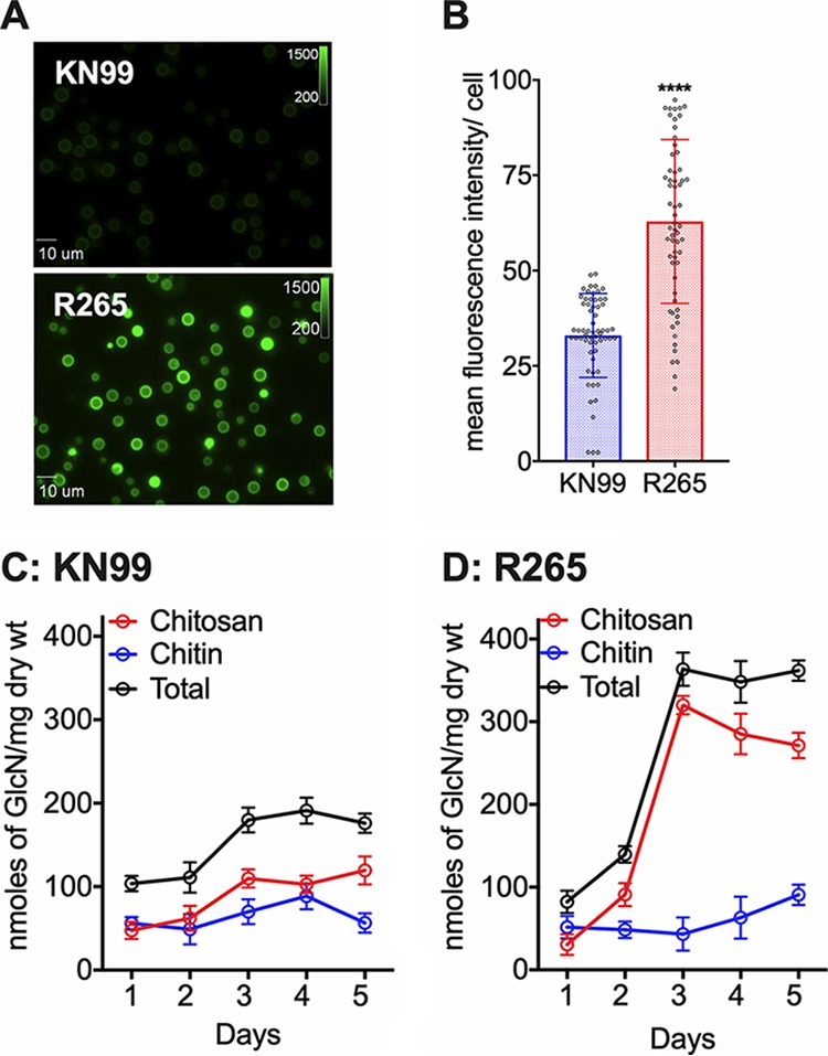
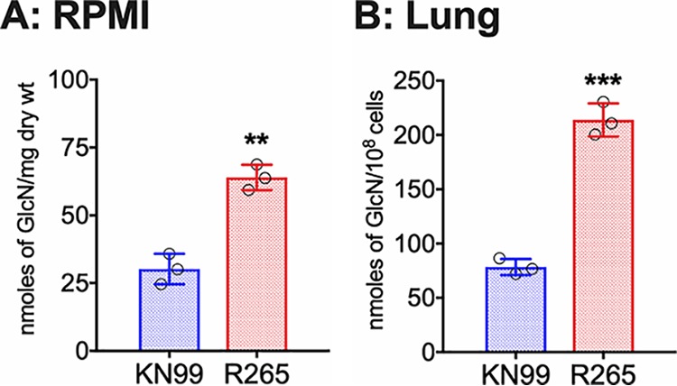
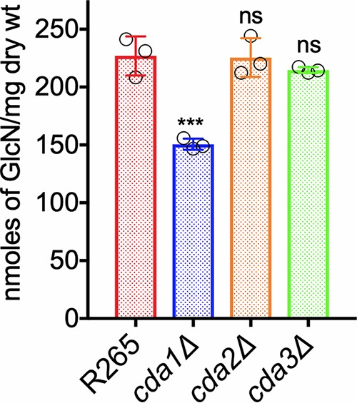
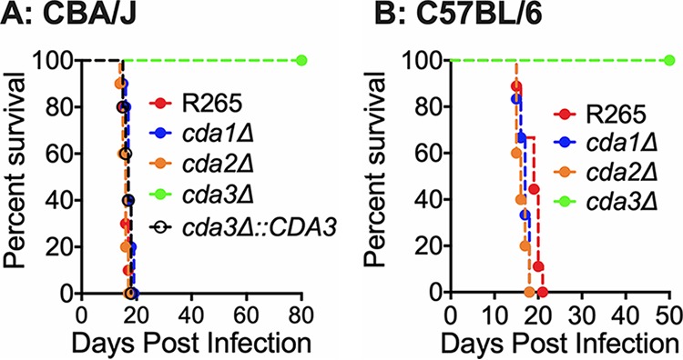
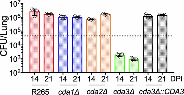
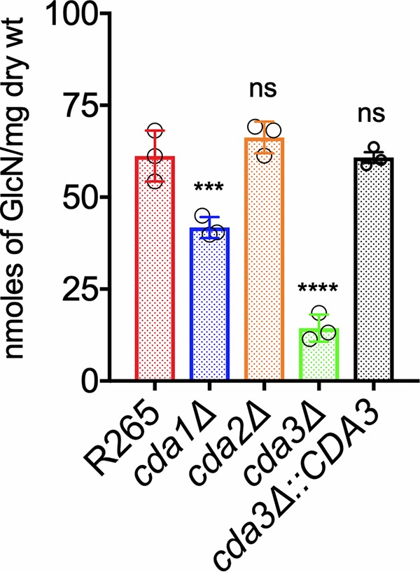
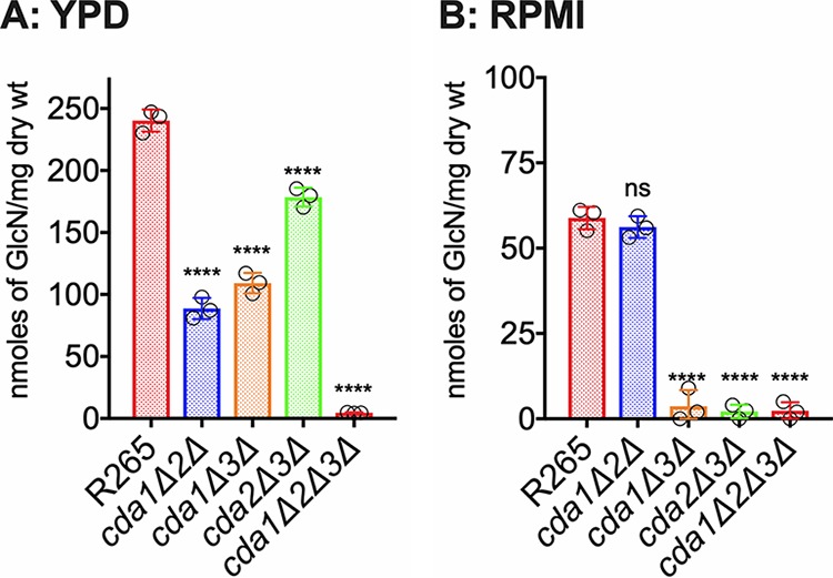
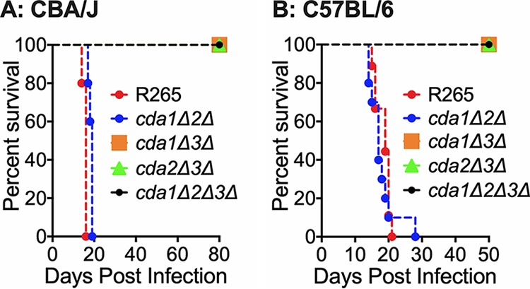
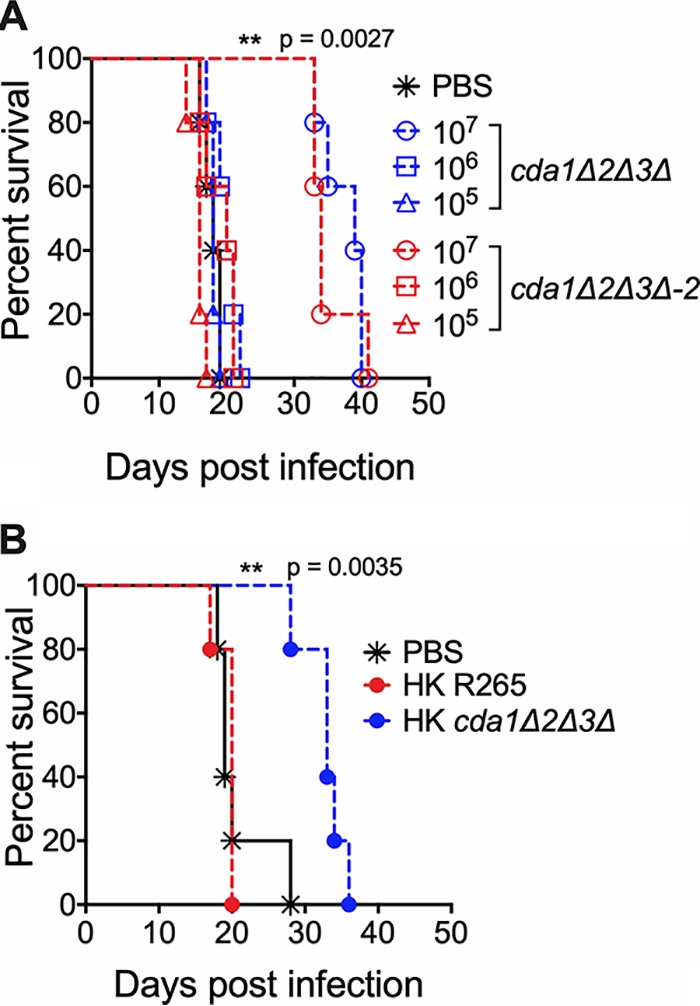
Cryptococcus gattii R265 is a hypervirulent fungal strain responsible for the recent outbreak of cryptococcosis in Vancouver Island of British Columbia in Canada. It differs significantly from Cryptococcus neoformans in its natural environment, its preferred site in the mammalian host, and its pathogenesis. Our previous studies of C. neoformans have shown that the presence of chitosan, the deacetylated form of chitin, in the cell wall attenuates inflammatory responses in the host, while its absence induces robust immune responses, which in turn facilitate clearance of the fungus and induces a protective response. The results of the present investigation reveal that the cell wall of C. gattii R265 contains a two- to threefold larger amount of chitosan than that of C. neoformans The genes responsible for the biosynthesis of chitosan are highly conserved in the R265 genome; the roles of the three chitin deacetylases (CDAs) have, however, been modified. To deduce their roles, single and double CDA deletion strains and a triple CDA deletion strain were constructed in a R265 background and were subjected to mammalian infection studies. Unlike C. neoformans where Cda1 has a discernible role in fungal pathogenesis, in strain R265, Cda3 is critical for virulence. Deletion of either CDA3 alone or in combination with another CDA (cda1Δ3Δ or cda2Δ3Δ) or both (cda1Δ2Δ3Δ) rendered the fungus avirulent and cleared from the infected host. Moreover, the cda1Δ2Δ3Δ strain of R265 induced a protective response to a subsequent infection with R265. These studies begin to illuminate the regulation of chitosan biosynthesis of C. gattii and its subsequent effect on fungal virulence.IMPORTANCE The fungal cell wall is an essential organelle whose components provide the first line of defense against host-induced antifungal activity. Chitosan is one of the carbohydrate polymers in the cell wall that significantly affects the outcome of host-pathogen interaction. Chitosan-deficient strains are avirulent, implicating chitosan as a critical virulence factor. C. gattii R265 is an important fungal pathogen of concern due to its ability to cause infections in individuals with no apparent immune dysfunction and an increasing geographical distribution. Characterization of the fungal cell wall and understanding the contribution of individual molecules of the cell wall matrix to fungal pathogenesis offer new therapeutic avenues for intervention. In this report, we show that the C. gattii R265 strain has evolved alternate regulation of chitosan biosynthesis under both laboratory growth conditions and during mammalian infection compared to that of C. neoformans.

Keywords: Cryptococcus gattii; R265; chitin; chitosan; chitosan regulation; protection; vaccine; virulence.


Figures

FIG 1
FIG 1
C. gattii R265 cells produce significantly larger amounts of chitosan in the cell wall compared to C. neoformans under YPD growth conditions. (A) Wild-type R265 and KN99 strains were grown in YPD for 5 days at 30°C and stained with Eosin Y to detect cell wall chitosan. Staining intensity was assessed using epifluorescence microscopy with identical exposures for all images. (B) Fluorescent levels for 60 individual cells (represented in panel A) were quantified using ImageJ (Fiji). The two-tailed unpaired t test with Welch’s correction was used to compare mean values of the wild type. Means represent the fluorescence intensity levels from three independent experiments (n = 3). ****, P < 0.0001. Error bars represent standard errors of the means. (C) Quantitative determination of cell wall chitosan and chitin of strain KN99 by the MBTH assay. Cells were grown in YPD for 1 to 5 days, collected, washed, and used for the assay. Data represents the averages of three biological experiments and are expressed as nanomoles of glucosamine per milligram (dry weight) of yeast cells. (D) Quantitative determination of cell wall chitosan and chitin of R265 were determined as in panel C.
FIG 2
FIG 2
C. gattii R265 cells produce significantly larger amount of chitosan in the cell wall compared to C. neoformans under host conditions. (A) Chitosan levels of strains grown in RPMI 1640 containing 10% FBS and 5% CO2 at 37°C for 5 days. Strains were grown in YPD for 2 days. Yeast cells were harvested, washed with PBS, inoculated at 500 cells/μl in RPMI 1640 containing 10% FBS, and incubated for 5 days at 37°C in the presence of 5% CO2. At the end of incubation, chitosan was measured by the MBTH assay and expressed as nanomoles of glucosamine per milligram (dry weight) of cells. Data represent the averages from three biological experiments. (B) Chitosan levels of strains growing in the murine lung. Mice (CBA/J) (three mice per group) were intranasally inoculated with 107 CFU of each strain. On day 7 postinfection (PI), the lungs were excised and homogenized, and the lung tissue was removed by alkaline extraction, leaving the fungal cells to be harvested, counted, and subjected to the MBTH assay. Data are expressed as nanomoles of glucosamine per 108 cells. Significant differences between the groups were compared by two-tailed unpaired t test with Welch’s correction. Error bars represent standard errors of the means. ***, P < 0.0062; **, P < 0.001.
FIG 3
FIG 3
The deletion of CDA1 in strain R265 displays a decrease in cell wall chitosan in cells grown in YPD. Chitosan levels of strains grown in YPD for 5 days were determined. The amount of chitosan in the cell wall of the strains was quantified by the MBTH assay. Data represent the averages from three biological experiments with two technical replicates and are expressed as nanomoles of glucosamine per milligram (dry weight) of yeast cells. Significant differences between the groups were compared by one-way ANOVA followed by Dunnett’s multiple-comparison test. ***, P < 0.0002 comparing wild-type R265 with any other strain. ns, not significant.
FIG 4
FIG 4
C. gattii cda3Δ displays severely attenuated virulence in CBA/J and C57BL/6 mouse models of infection. (A) CBA/J mice (6 to 8 weeks old, female) were infected intranasally with 105 CFU of each strain. Survival of the animals was recorded as mortality of mice for 80 days PI. Mice that lost 20% of the body weight at the time of inoculation were considered ill and sacrificed. Data are representative of two independent experiments with five animals for each strain. (B) C57BL/6 mice (4 to 6 weeks old, female) were infected with 104 CFU of each strain by intratracheal inoculation. Survival of the animals was recorded as mortality of mice for 50 days PI. Mice that lost 20% of the body weight at the time of inoculation were considered ill and sacrificed. Data are representative of one experiment with 10 animals for each strain. Virulence was determined using Mantel-Cox curve comparison with statistical significance determined by log rank test. P < 0.0001 comparing strain KN99 with cda3Δ strain.
FIG 5
FIG 5
There is a slow but gradual clearance of C. gattii cda3Δ in CBA/J mice. Fungal burden in the lungs of CBA/J mice infected with indicated strains at different days postinfection (DPI). Data are from three mice per group at each time point. The dotted line indicates the CFU of the initial inoculum for each strain.
FIG 6
FIG 6
C. gattii Cda3 plays a major role in the synthesis of chitosan under host infection conditions. Chitosan levels of strains grown in RPMI 1640 containing 10% FBS and 5% CO2 at 37°C for 5 days. The indicated strains were grown in YPD for 48 h. Yeast cells were harvested, washed with PBS, and inoculated at 500 cells/μl in RPMI 1640 containing 10% FBS and incubated for 5 days at 37°C in the presence of 5% CO2. At the end of incubation, chitosan was measured by the MBTH assay and expressed as nanomoles of glucosamine per milligram (dry weight) of cells. Data represent the averages from three biological experiments. Significant differences between the groups were compared by one-way ANOVA, followed by Dunnett’s multiple-comparison test (***, P < 0.0002, *, P < 0.0280 comparing KN99 with any other strain; ns, not significant).
FIG 7
FIG 7
The deletion of C. gattii CDA1, CDA2, and CDA3 results in a strain that is completely chitosan deficient. C. gattii Cda1 in combination with Cda2 or Cda3 plays a major role in chitosan synthesis in vegetative growing conditions. While C. gattii Cda3 in combination with Cda1 or Cda2 results in a stain that is completely chitosan-deficient. (A) Chitosan levels of strains grown in YPD. The indicated strains were grown in YPD for 5 days. The amount of chitosan in the cell wall of the strains was quantified by the MBTH assay. Data are the averages for three biological experiments and are expressed as nanomoles of glucosamine per milligram (dry weight) of yeast cells. (B) Chitosan levels of strains grown in RPMI 1640 containing 10% FBS and 5% CO2 at 37°C for 5 days. The indicated strains were grown in YPD for 48 h. Yeast cells were harvested, washed with PBS, and inoculated at 500 cells/μl in RPMI 1640 containing 10% FBS and incubated for 5 days at 37°C in the presence of 5% CO2. At the end of incubation, chitosan was measured by the MBTH assay and expressed as nanomoles of glucosamine per milligram (dry weight) of cells. Data represent the averages for three biological experiments. Significant differences between the groups were compared by one-way ANOVA, followed by Dunnett’s multiple-comparison test (****, P < 0.0001, comparing KN99 with any other strain; ns, not significant).
FIG 8
FIG 8
Deletion of C. gattii CDA3 in combination with any of the other two CDAs results in severe attenuation of virulence in CBA/J and C57BL/6 mouse models of infection. (A) CBA/J mice (6 to 8 weeks old, female) were infected intranasally with 105 CFU of each strain. Survival of the animals was recorded as mortality of mice for 80 days postinfection (PI). Mice that lost 20% of the body weight at the time of inoculation were considered ill and sacrificed. Data are representative of two independent experiments with five animals for each strain. (B) C57BL/6 mice (4 to 6 weeks old, female) were infected intratracheally with 104 CFU of each strain. Survival of the animals was recorded as mortality of mice for 50 days PI. Mice that lost 20% of the body weight at the time of inoculation were considered ill and sacrificed.
FIG 9
FIG 9
Vaccination of CBA/J mice with 107 CFU of live or heat-killed (HK) cda1Δ2Δ3Δ cells conferred attenuated protective immunity to subsequent infection with wild-type R265 C. gattii cells. (A) Mice were immunized with either 107, 106, or 105 live CFU of cda1Δ2Δ3Δ cells through inhalation. PBS-inoculated mice served as control. Animals were left for 40 days to resolve the infection. Subsequently, both groups of mice were challenged with 50,000 CFU of virulent R265 cells. Virulence was recorded as mortality of mice. Mice that lost 20% of starting body weight at the time of inoculation were considered to be moribund and sacrificed. The percentage of mice that survived was plotted against the day postinfection. Each survival curve is the average of two independent experiments that had five mice per experimental group. (B) Mice were immunized with an inoculum of HK cells with a dose equivalent to 107 CFU of either the wild-type R265 or the cda1Δ2Δ3Δ strain. Control mice were inoculated with PBS. After 40 days, mice were challenged with 50,000 CFU of wild-type R265 cells. Survival of the animals was recorded as described above. The data shown are from a single experiment with five mice per experimental group. A second experiment showed similar results.

References

    1. Spickler AR. 2013. Cryptococcosis. The Center for Food Security and Public Health, Iowa State University, Ames, Iowa.
    1. Barnett JA. 2010. A history of research on yeasts 14: medical yeasts part 2, Cryptococcus neoformans. Yeast 27:875–904. doi:10.1002/yea.1786. - DOI - PubMed
    1. Rajasingham R, Smith RM, Park BJ, Jarvis JN, Govender NP, Chiller TM, Denning DW, Loyse A, Boulware DR. 2017. Global burden of disease of HIV-associated cryptococcal meningitis: an updated analysis. Lancet Infect Dis 17:873–881. doi:10.1016/S1473-3099(17)30243-8. - DOI - PMC - PubMed
    1. Idnurm A, Bahn YS, Nielsen K, Lin X, Fraser JA, Heitman J. 2005. Deciphering the model pathogenic fungus Cryptococcus neoformans. Nat Rev Microbiol 3:753–764. doi:10.1038/nrmicro1245. - DOI - PubMed
    1. Kronstad JW, Attarian R, Cadieux B, Choi J, D’Souza CA, Griffiths EJ, Geddes JMH, Hu G, Jung WH, Kretschmer M, Saikia S, Wang J. 2011. Expanding fungal pathogenesis: Cryptococcus breaks out of the opportunistic box. Nat Rev Microbiol 9:193–203. doi:10.1038/nrmicro2522. - DOI - PMC - PubMed

Publication types

MeSH terms

LinkOut - more resources