. 2019 Dec 12;94(1):e01174-19.
doi: 10.1128/JVI.01174-19. Print 2019 Dec 12.

Infrequent HIV Infection of Circulating Monocytes during Antiretroviral Therapy

Affiliations

Infrequent HIV Infection of Circulating Monocytes during Antiretroviral Therapy

Marta Massanella et al. J Virol. .

Abstract

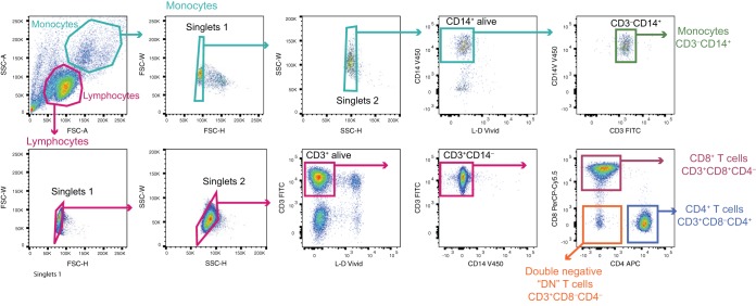
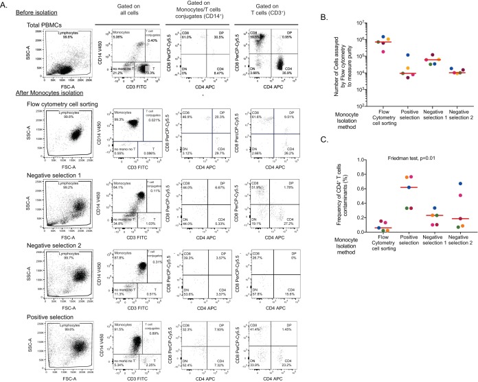
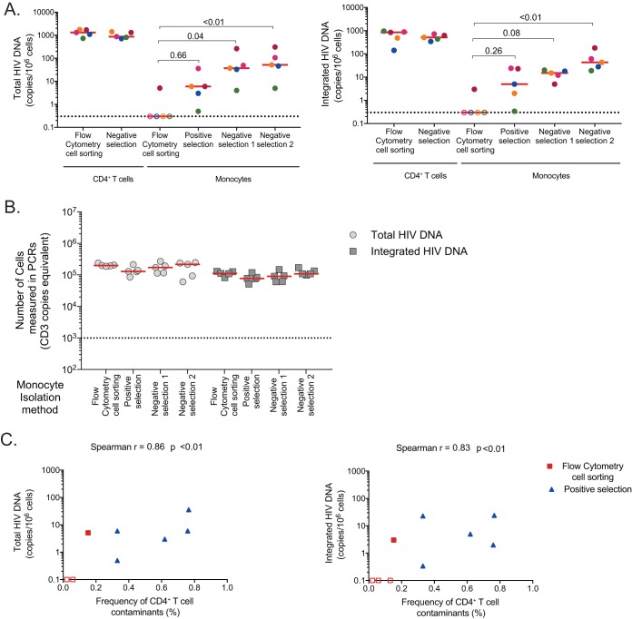
Whereas human immunodeficiency virus (HIV) persists in tissue macrophages during antiretroviral therapy (ART), the role of circulating monocytes as HIV reservoirs remains controversial. Three magnetic bead selection methods and flow cytometry cell sorting were compared for their capacity to yield pure CD14+ monocyte populations. Cell sorting by flow cytometry provided the purest population of monocytes (median CD4+ T-cell contamination, 0.06%), and the levels of CD4+ T-cell contamination were positively correlated with the levels of integrated HIV DNA in the monocyte populations. Using cell sorting by flow cytometry, we assessed longitudinally the infection of monocytes and other cell subsets in a cohort of 29 Thai HIV-infected individuals. Low levels of HIV DNA were detected in a minority of monocyte fractions obtained before and after 1 year of ART (27% and 33%, respectively), whereas HIV DNA was readily detected in CD4+ T cells from all samples. Additional samples (2 to 5 years of ART) were obtained from 5 individuals in whom monocyte infection was previously detected. Whereas CD4+ T cells were infected at high levels at all time points, monocyte infection was inconsistent and absent in at least one longitudinal sample from 4/5 individuals. Our results indicate that infection of monocytes is infrequent and highlight the importance of using flow cytometry cell sorting to minimize contamination by CD4+ T cells.IMPORTANCE The role of circulating monocytes as persistent HIV reservoirs during ART is still controversial. Several studies have reported persistent infection of monocytes in virally suppressed individuals; however, others failed to detect HIV in this subset. These discrepancies are likely explained by the diversity of the methods used to isolate monocytes and to detect HIV infection. In this study, we show that only flow cytometry cell sorting yields a highly pure population of monocytes largely devoid of CD4 contaminants. Using this approach in a longitudinal cohort of HIV-infected individuals before and during ART, we demonstrate that HIV is rarely found in monocytes from untreated and treated HIV-infected individuals. This study highlights the importance of using methods that yield highly pure populations of cells as flow cytometry cell sorting to minimize and control for CD4+ T-cell contamination.

Keywords: HIV reservoir; integrated HIV DNA; monocytes.


Figures

FIG 1
FIG 1
Gating strategy for cell sorting. Flow cytometry dot plots showing the gating strategy used to isolate CD4+ T cells (CD3+ CD4+ CD8), CD8+ T cells (CD3+ CD4 CD8+), double-negative (DN) cells (CD3+ CD4 CD8), and monocytes (CD3 CD14+).
FIG 2
FIG 2
Assessment of monocyte infection following different methods of isolation. Three magnetic bead selection methods (one positive and two negative selection) and flow cytometry cell sorting of CD14+ cells were used to isolate monocytes. (A) Gating strategy for purity analysis. Flow cytometry dot plots showing the cellular distribution of PBMCs (upper lane) and enriched monocytes following isolation by cell sorting or three magnetic bead selection methods (positive and negative selection). A sample from one representative donor is shown. SSC, side scatter; FITC, fluorescein isothiocyanate; APC, antigen-presenting cell; FSC, forward scatter. (B) Number of monocytes analyzed by flow cytometry. (C) Frequency of CD4+ T-cell contaminants after monocyte enrichment determined by flow cytometry in postenrichment samples.
FIG 3
FIG 3
Assessment of monocyte infection following different methods of isolation. (A) Frequency of total (left panel) and integrated HIV DNA (right panel) in enriched CD4+ T cells isolated by flow cytometry cell sorting and negative selection, as well as in monocytes isolated using the four methods described for Fig. 2. (B) Number of monocytes analyzed by total and integrated HIV PCR for each sample. C) Correlation between the levels of total (left) or integrated (right) HIV DNA measured in the monocyte fractions and the frequency of residual CD4+ T cells in this subset. Monocytes obtained by flow cytometry cell sorting (in red) and positive magnetic bead selection (in blue) are shown. Due to the high monocyte autofluorescence detected after enrichment with the two negative magnetic bead selection methods, these were excluded from the analysis.
FIG 4
FIG 4
Levels of HIV DNA in circulating cell-sorted subsets after correction for CD4+ T-cell contamination. PBMCs, as well as isolated CD4+, CD8+, double-negative (DN) T cells (CD8 CD4), and monocytes were sorted by flow cytometry from the blood of 29 Thai individuals and subjected to integrated HIV DNA quantification. (A) Number of cells analyzed as measured by PCR in PBMCs and in each sorted subset are indicated. Only samples in which at least 10,000 cells (dashed horizontal line) were assayed were included in the succeeding analysis. LOD, limit of detection. (B) Levels of integrated HIV DNA in PBMCs, CD4+ T cells, monocytes, and DN and CD8+ T cells before and after 1 year of ART. Numbers of samples in which HIV DNA was detected (and the corresponding percentages) are indicated. Each individual is color-coded, and horizontal bars indicate median values. (C) Frequencies of residual CD4+ T-cell in CD8+ T-cell, DN, and monocyte enriched populations post-cell sorting. The numbers of samples with measurable residual CD4+ T cells (and the corresponding percentages) are indicated. (D) The levels of HIV DNA in monocytes, DN T cells, and CD8+ T cells were adjusted for CD4+ T-cell contamination. The numbers of samples in which HIV DNA was detected (and the corresponding percentages) are indicated. Samples from each participant are color-coded, and horizontal bars indicate median values. (E) Integrated HIV DNA levels from paired samples obtained at baseline (before ART initiation) and after 1 year of ART in CD4+ T cells, monocytes, DN T cells, and CD8+ T cells. P values were obtained from the Wilcoxon matched-pair signed-rank test. (F) Correlation between the levels of integrated HIV DNA at baseline and after 1 year of ART in CD4+ T cells. (G) Correlations between the frequency of CD4+ T cells harboring integrated HIV DNA and the levels of integrated HIV DNA measured in monocytes (upper left), DN T cells (upper middle), and CD8 T cells (upper right). Similar correlations were repeated after adjusting for CD4+ T-cell contamination (bottom row). (F and G) P values were obtained using the Spearman test. (H) Pie charts representing the contribution of each subset (CD4+ T cells [blue], monocytes [red], DN T cells [green], and CD8+ T cells [yellow]) to the total pool of cells harboring integrated HIV DNA at baseline (before ART, left) and after 1 year on ART (right).
FIG 5
FIG 5
Longitudinal assessment of HIV infection in CD4+ T cells and monocytes. (A) Number of cells at each time point measured by PCR in sorted CD4+ T cells (left) and monocytes (right) in five individuals in whom HIV DNA was previously detected (either at baseline or after 1 year of ART) in the monocyte fraction. (B) Levels of integrated HIV DNA in CD4+ T cells (left) and monocytes (right) were assessed longitudinally in five individuals in whom HIV DNA was previously detected (either at baseline or after 1 year of ART) in the monocyte fraction. The levels of HIV DNA in monocytes were adjusted for CD4+ T-cell contamination. Only samples in which a minimum of 10,000 cells were analyzed were included. Each individual’s data are color-coded.

References

    1. Murray JM, Zaunders JJ, McBride KL, Xu Y, Bailey M, Suzuki K, Cooper DA, Emery S, Kelleher AD, Koelsch KK, PINT Study Team . 2014. HIV DNA subspecies persist in both activated and resting memory CD4+ T cells during antiretroviral therapy. J Virol 88:3516–3526. doi: 10.1128/JVI.03331-13. - DOI - PMC - PubMed
    1. Garrigue I, Pellegrin I, Hoen B, Dumon B, Harzic M, Schrive MH, Séréni D, Fleury H. 2000. Cell-associated HIV-1-DNA quantitation after highly active antiretroviral therapy-treated primary infection in patients with persistently undetectable plasma HIV-1 RNA. AIDS 14:2851–2855. doi: 10.1097/00002030-200012220-00006. - DOI - PubMed
    1. Besson GJ, Lalama CM, Bosch RJ, Gandhi RT, Bedison MA, Aga E, Riddler SA, McMahon DK, Hong F, Mellors JW. 2014. HIV-1 DNA decay dynamics in blood during more than a decade of suppressive antiretroviral therapy. Clin Infect Dis 59:1312–1321. doi: 10.1093/cid/ciu585. - DOI - PMC - PubMed
    1. Wong JK, Hezareh M, Günthard HF, Havlir DV, Ignacio CC, Spina CA, Richman DD. 1997. Recovery of replication-competent HIV despite prolonged suppression of plasma viremia. Science 278:1291–1295. doi: 10.1126/science.278.5341.1291. - DOI - PubMed
    1. Finzi D, Hermankova M, Pierson T, Carruth LM, Buck C, Chaisson RE, Quinn TC, Chadwick K, Margolick J, Brookmeyer R, Gallant J, Markowitz M, Ho DD, Richman DD, Siliciano RF. 1997. Identification of a reservoir for HIV-1 in patients on highly active antiretroviral therapy. Science 278:1295–1300. doi: 10.1126/science.278.5341.1295. - DOI - PubMed

Publication types

MeSH terms