. 2019 Dec 12;94(1):e01534-19.
doi: 10.1128/JVI.01534-19. Print 2019 Dec 12.

Isolation of Yasminevirus, the First Member of Klosneuvirinae Isolated in Coculture with Vermamoeba vermiformis, Demonstrates an Extended Arsenal of Translational Apparatus Components

Affiliations

Isolation of Yasminevirus, the First Member of Klosneuvirinae Isolated in Coculture with Vermamoeba vermiformis, Demonstrates an Extended Arsenal of Translational Apparatus Components

Leena Hussein Bajrai et al. J Virol. .

Abstract

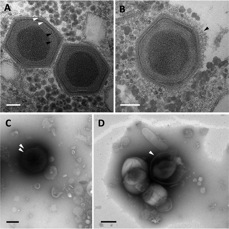
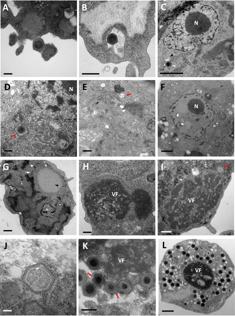
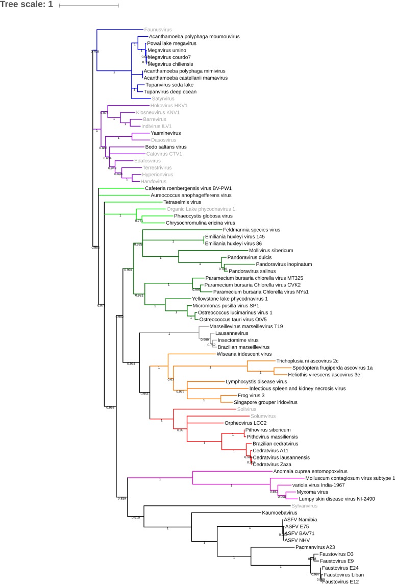
The family of giant viruses is still expanding, and evidence of a translational machinery is emerging in the virosphere. The Klosneuvirinae group of giant viruses was first reconstructed from in silico studies, and then a unique member was isolated, Bodo saltans virus. Here we describe the isolation of a new member in this group using coculture with the free-living amoeba Vermamoeba vermiformis This giant virus, called Yasminevirus, has a 2.1-Mb linear double-stranded DNA genome encoding 1,541 candidate proteins, with a GC content estimated at 40.2%. Yasminevirus possesses a nearly complete translational machinery, with a set of 70 tRNAs associated with 45 codons and recognizing 20 amino acids (aa), 20 aminoacyl-tRNA synthetases (aaRSs) recognizing 20 aa, as well as several translation factors and elongation factors. At the genome scale, evolutionary analyses placed this virus in the Klosneuvirinae group of giant viruses. Rhizome analysis demonstrated that the genome of Yasminevirus is mosaic, with ∼34% of genes having their closest homologues in other viruses, followed by ∼13.2% in Eukaryota, ∼7.2% in Bacteria, and less than 1% in Archaea Among giant virus sequences, Yasminevirus shared 87% of viral hits with Klosneuvirinae. This description of Yasminevirus sheds light on the Klosneuvirinae group in a captivating quest to understand the evolution and diversity of giant viruses.IMPORTANCE Yasminevirus is an icosahedral double-stranded DNA virus isolated from sewage water by amoeba coculture. Here its structure and replicative cycle in the amoeba Vermamoeba vermiformis are described and genomic and evolutionary studies are reported. This virus belongs to the Klosneuvirinae group of giant viruses, representing the second isolated and cultivated giant virus in this group, and is the first isolated using a coculture procedure. Extended translational machinery pointed to Yasminevirus among the quasiautonomous giant viruses with the most complete translational apparatus of the known virosphere.

Keywords: Klosneuvirinae; NCLDV; amoeba; giant virus; translation.


Figures

FIG 1
FIG 1
Morphologic features and ultrastructure of Yasminevirus mature particles. (A and B) Observation of mature Yasminevirus particles by transmission electron microscopy. (A) Transmission electron microscopy image of Yasminevirus particles highlighting the multiple inner layers surrounding the viral core genome. From the inside to the outside, two putative thin membranes, similar to those observed in Bodo saltans virus (black arrowheads), and the viral capsid composed of two visible layers (white arrowheads) can be observed. Scale bar, 100 nm. (B) The outer capsid of Yasminevirus is surrounded by a thin layer of fibrils (arrowhead). Scale bar, 100 nm. (C and D) Observation of mature Yasminevirus particles with negative-staining electron microscopy. Two distinct aspects of virus particles can be observed in a culture supernatant of Yasminevirus. (C) Virus particles with an intact double capsid (arrowhead). (D) Particles with broken capsids, giving rise to a pacmanvirus-like morphology (arrowhead).
FIG 2
FIG 2
Characterization of the Yasminevirus replication cycle with transmission electron microscopy. (A) Yasminevirus particle attachment to the Vermamoeba vermiformis cytoplasmic membrane before its internalization by phagocytosis. Scale bar, 200 nm. (B) Virus particles inside the phagosome. Scale bar, 500 nm. (C) Once in the cytoplasm, the virus seems to escape the phagosome and loses its outer capsid (black arrowhead). The Yasminevirus particle is indicated by a white arrowhead. Scale bar, 1 μm. N, nucleus. (D) The virus particle loses its internal capsid before the genome release step. Scale bar, 200 nm. (E) Empty particles after the release of the viral content into the amoeba cytoplasm (arrow). Scale bar, 500 nm. (F) The eclipse phase is marked by a remodeling of the cell nucleus by the appearance of cleared areas inside the nucleus (arrowheads). Scale bar, 1 μm. (G) The early stages of infection are then characterized by the appearance of a bright area in the cytoplasm of the host cell, which represents the early virus factory (black arrowhead). The mitochondria recruited around the virus factory are highlighted with white arrowheads. Scale bar, 1 μm. (H) At a later stage of infection, the virus factory (VF) starts to appear denser and occupies a larger part of the host cell cytoplasm. Scale bar, 500 nm. (I) Assembly of virions, the first virus progeny (arrow). Scale bar, 500 nm. (J) Immature virion on which the double proteinaceous capsid is completely assembled (arrowheads). Scale bar, 100 nm. (K) In some cases, genome acquisition could occur before the complete assembly of the virion (arrows). Viral particles that have already acquired their genomes have an electron-dense region at the center of the capsids. Scale bar, 400 nm. (L) At the end of the viral cycle, mature virions occupy the whole host cell cytoplasm up until host cell lysis. Scale bar, 1 μm.
FIG 3
FIG 3
Graphical circular map of the genome of Yasminevirus. From outside to center: scaffolds (orange/brown), genes on the forward strand (dark brown), genes on the reverse strand (blue), tRNA (green), and GC skew (light green and purple).
FIG 4
FIG 4
Translation system components in Yasminevirus. tRNA, aminoacyl-tRNA synthetase (aaRS), and translation factors (TF) are compared in the Klosneuvirinae group and in Tupanvirus.
FIG 5
FIG 5
The rhizome of Yasminevirus. Viruses are depicted in red, Archaea in pink, Eukaryota in blue, and Bacteria in green. ORFans are depicted in orange. All protein sequences were used as queries in a BLASTp search against the nonredundant (nr) protein database from NCBI. BLAST results were filtered to keep the best hits, and taxonomic affiliation was retrieved from the NCBI database. The best hit was selected and integrated in a circular genomic data image (37).
FIG 6
FIG 6
Phylogenetic tree of Yasminevirus based on DNA polymerase B proteins of nucleocytoplasmic large DNA viruses (NCLDVs). Colors have been assigned for the different virus groups as follows: blue for Mimivirus and extended Mimiviridae; green for pandoraviruses, Mollivirus sibericum, and Phycodnaviridae; black for groups of Asfarviridae, faustoviruses, pacmanvirus, and kaumoebavirus; gray for Marseilleviridae; red for orpheovirus, cedratvirus, and pithoviruses; and purple for asco-iridoviridae.
FIG 7
FIG 7
Orthologous genes shared between Yasminevirus and other members within Klosneuvirinae.

References

    1. La Scola B, Audic S, Robert C, Jungang L, De Lamballerie X, Drancourt M, Birtles R, Claverie JM, Raoult D. 2003. A giant virus in amoebae. Science 299:2033. doi:10.1126/science.1081867. - DOI - PubMed
    1. Colson P, La Scola B, Levasseur A, Caetano-Anollés G, Raoult D. 2017. Mimivirus: leading the way in the discovery of giant viruses of amoebae. Nat Rev Microbiol 15:243–254. doi:10.1038/nrmicro.2016.197. - DOI - PMC - PubMed
    1. Forterre P. 2017. Viruses in the 21st century: from the curiosity-driven discovery of giant viruses to new concepts and definition of life. Clin Infect Dis 65:S74–S79. doi:10.1093/cid/cix349. - DOI - PubMed
    1. Claverie JM, Abergel C. 2016. Giant viruses: update, enigmas, controversies and perspectives. Med Sci (Paris) 32:1087–1096. doi:10.1051/medsci/20163212012. - DOI - PubMed
    1. Levasseur A, Andreani J, Delerce J, Bou Khalil J, Robert C, La Scola B, Raoult D. 2016. Comparison of a modern and fossil pithovirus reveals its genetic conservation and evolution. Genome Biol Evol 8:2333–2339. doi:10.1093/gbe/evw153. - DOI - PMC - PubMed

Publication types

MeSH terms

LinkOut - more resources