. 2019 Dec 12;94(1):e01623-19.
doi: 10.1128/JVI.01623-19. Print 2019 Dec 12.

Yellow Fever Virus Reemergence and Spread in Southeast Brazil, 2016-2019

Affiliations

Yellow Fever Virus Reemergence and Spread in Southeast Brazil, 2016-2019

Marta Giovanetti et al. J Virol. .

Erratum in

  • Correction for Giovanetti et al., "Yellow Fever Virus Reemergence and Spread in Southeast Brazil, 2016-2019".
    Giovanetti M, Lima de Mendonça MC, Fonseca V, Mares-Guia MA, Fabri A, Xavier J, Goes de Jesus J, Gräf T, Damasceno Dos Santos Rodrigues C, Cardoso Dos Santos C, Alves Sampaio S, Lowen Levy Chalhoub F, de Bruycker Nogueira F, Theze J, Pecego Martins Romano A, Garkauskas Ramos D, Luiz de Abreu A, Kleber Oliveira W, do Carmo Said RF, Campelo de Alburque CF, de Oliveira T, Fernandes CA, Ferreira Aguiar S, Chieppe A, Carvalho Sequeira P, Rodrigues Faria N, Venâncio Cunha R, Alcantara LCJ, Bispo de Filippis AM. Giovanetti M, et al. J Virol. 2020 May 18;94(11):e02008-19. doi: 10.1128/JVI.02008-19. Print 2020 May 18. J Virol. 2020. PMID: 32423967 Free PMC article. No abstract available.

Abstract

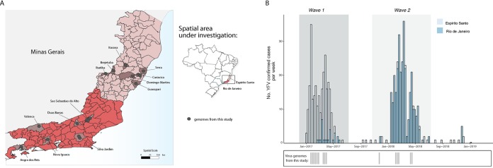
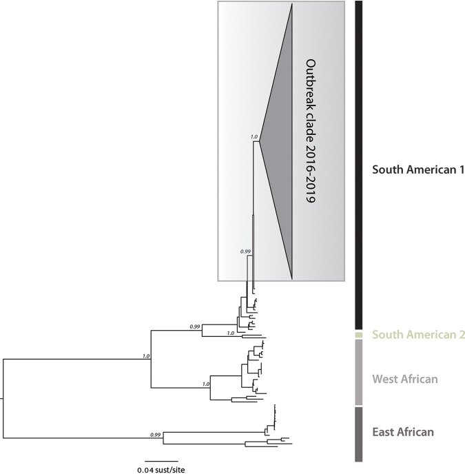
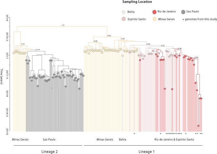
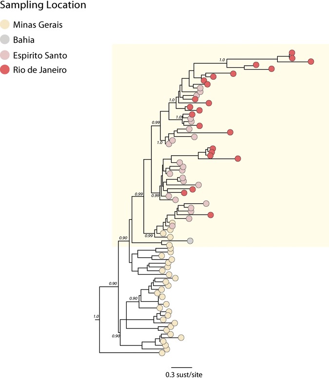
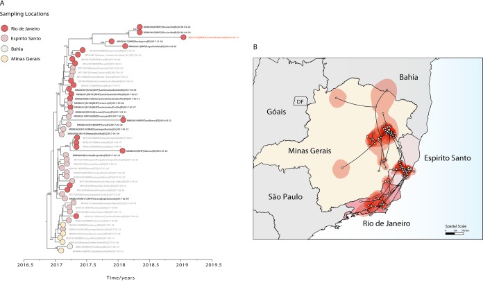
The recent reemergence of yellow fever virus (YFV) in Brazil has raised serious concerns due to the rapid dissemination of the virus in the southeastern region. To better understand YFV genetic diversity and dynamics during the recent outbreak in southeastern Brazil, we generated 18 complete and nearly complete genomes from the peak of the epidemic curve from nonhuman primates (NHPs) and human infected cases across the Espírito Santo and Rio de Janeiro states. Genomic sequencing of 18 YFV genomes revealed the estimated timing, source, and likely routes of yellow fever virus transmission and dispersion during one of the largest outbreaks ever registered in Brazil. We showed that during the recent epidemic, YFV was reintroduced from Minas Gerais to the Espírito Santo and Rio de Janeiro states multiple times between 2016 and 2019. The analysis of data from portable sequencing could identify the corridor of spread of YFV. These findings reinforce the idea that continued genomic surveillance strategies can provide information on virus genetic diversity and transmission dynamics that might assist in understanding arbovirus epidemics.IMPORTANCE Arbovirus infections in Brazil, including yellow fever, dengue, zika, and chikungunya, result in considerable morbidity and mortality and are pressing public health concerns. However, our understanding of these outbreaks is hampered by the limited availability of genomic data. In this study, we investigated the genetic diversity and spatial distribution of YFV during the current outbreak by analyzing genomic data from areas in southeastern Brazil not covered by other previous studies. To gain insights into the routes of YFV introduction and dispersion, we tracked the virus by sequencing YFV genomes sampled from nonhuman primates and infected patients from the southeastern region. Our study provides an understanding of how YFV initiates transmission in new Brazilian regions and illustrates that genomics in the field can augment traditional approaches to infectious disease surveillance and control.

Keywords: Southeast Brazil; genomic surveillance; outbreak; outbreak response; yellow fever.


Figures

FIG 1
FIG 1
Spatial and temporal distribution of YF cases from the Espírito Santo and Rio de Janeiro states during 2017 and 2019. (A) Map of the states of Espírito Santo (ES) and Rio de Janeiro (RJ), located in the southeastern region of Brazil and its municipalities. Circles indicate where samples from this study were collected. (B) Time series of human (H) and nonhuman primate YFV cases in ES and RJ states confirmed by serology, reverse transcription quantitative PCR (RT-qPCR), or virus isolation. Below, the dates of sample collection of the virus genomes generated in this study are shown in gray bars.
FIG 2
FIG 2
Molecular phylogenetics of the Brazilian YFV epidemic. Maximum likelihood phylogeny of complete YFV genomes showing the outbreak clade (gray triangle) within the South American I (SA1) genotype. The scale bar is in units of substitutions per site (sust/site).
FIG 3
FIG 3
Time-scaled phylogenetic tree of the current YF epidemic in Brazil. Molecular clock phylogeny obtained by combining the 18 new YFV complete genomes generated here (starred tips) plus publicly available data (n = 137) of the YFV 2016 to 2019 epidemic in Brazil (11, 12, 17, 22, 23). Numbers in nodes represent clade posterior probability of >0.90. Branch colors represent different sampling locations.
FIG 4
FIG 4
Molecular clock phylogeny including the clade comprising the new isolates plus all the YFV strains from the 2017 to 2019 outbreak belonging to the SA1 lineage 1 clade. Numbers along branches represent clade posterior probability of >0.90. Colors represent different locations.
FIG 5
FIG 5
Spatiotemporal dynamics of YFV SA1 lineage 1. (A) Molecular clock phylogeny including the clade comprising the 2017 to 2019 YFV strains from Minas Gerais, Bahia, Espírito Santo, and Rio de Janeiro states belonging to SA1 lineage 1. Numbers along branches represent clade posterior probability of >0.90. YFV isolates from Casimiro de Abreu, sampled in January 2019, are highlighted in red. Colors represent different locations. (B) Reconstructed spatiotemporal continuous diffusion of the YFV SA1 lineage 1 outbreak clade. Phylogenetic branches are mapped in space according to the location of phylogenetic nodes (circles). Lines show the cross-state movement of the virus from Minas Gerais followed by movement to the states of Espírito Santo and Rio de Janeiro. Shaded regions show 95% credible regions of internal nodes.

Similar articles

Cited by

References

    1. Barrett AD, Monath TP. 2003. Epidemiology and ecology of yellow fever virus. Adv Virus Res 61:291–315. doi:10.1016/S0065-3527(03)61007-9. - DOI - PubMed
    1. Monath TP, Vasconcelos PF. 2015. Yellow fever. J Clin Virol 64:160–173. doi:10.1016/j.jcv.2014.08.030. - DOI - PubMed
    1. Bryant JE, Holmes EC, Barrett AD. 2007. Out of Africa: a molecular perspective on the introduction of yellow fever virus into the Americas. PLoS Pathog 3:e75. doi:10.1371/journal.ppat.0030075. - DOI - PMC - PubMed
    1. Mutebi JP, Wang H, Li L, Bryant JE, Barrett AD. 2001. Phylogenetic and evolutionary relationships among yellow fever virus isolates in Africa. J Virol 75:6999–7008. doi:10.1128/JVI.75.15.6999-7008.2001. - DOI - PMC - PubMed
    1. Nunes MR, Palacios G, Cardoso JF, Martins LC, Sousa EC Jr, de Lima CP, Medeiros DB, Savji N, Desai A, Rodrigues SG, Carvalho VL, Lipkin WI, Vasconcelos PF. 2012. Genomic and phylogenetic characterization of Brazilian yellow fever virus strains. J Virol 86:13263–13271. doi:10.1128/JVI.00565-12. - DOI - PMC - PubMed

Publication types

MeSH terms

LinkOut - more resources