

Multiscale three-dimensional surface reconstruction and surface roughness of porcine left anterior descending coronary arteries
- PMID: 31598314
- PMCID: PMC6774942
- DOI: 10.1098/rsos.190915
Multiscale three-dimensional surface reconstruction and surface roughness of porcine left anterior descending coronary arteries
Abstract
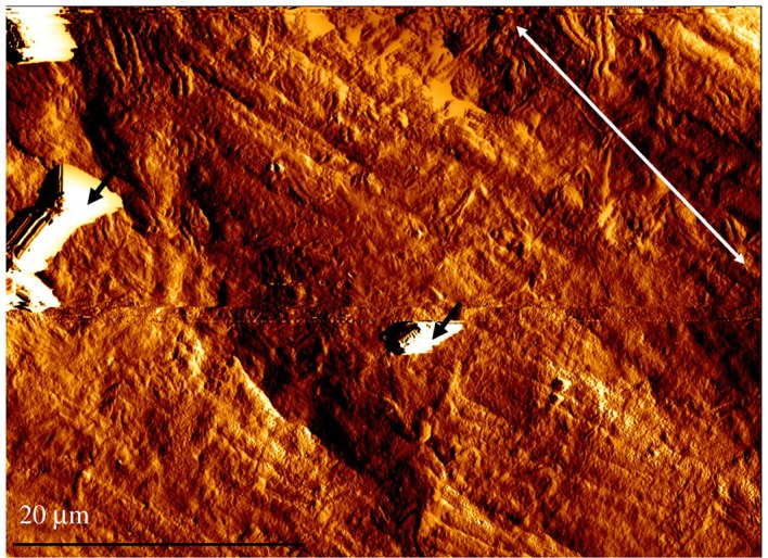
The aim of this study was to investigate the multiscale surface roughness characteristics of coronary arteries, to aid in the development of novel biomaterials and bioinspired medical devices. Porcine left anterior descending coronary arteries were dissected ex vivo, and specimens were chemically fixed and dehydrated for testing. Surface roughness was calculated from three-dimensional reconstructed surface images obtained by optical, scanning electron and atomic force microscopy, ranging in magnification from 10× to 5500×. Circumferential surface roughness decreased with magnification, and microscopy type was found to influence surface roughness values. Longitudinal surface roughness was not affected by magnification or microscopy types within the parameters of this study. This study found that coronary arteries exhibit multiscale characteristics. It also highlights the importance of ensuring consistent microscopy parameters to provide comparable surface roughness values.
Keywords: atomic force microscopy; coronary arteries; endothelium; multiscale; scanning electron microscopy; surface roughness.
© 2019 The Authors.
Conflict of interest statement
The authors declare that they have no competing interests.
Figures


Similar articles
-
Dynamic Viscoelasticity and Surface Properties of Porcine Left Anterior Descending Coronary Arteries.Cardiovasc Eng Technol. 2017 Mar;8(1):41-56. doi: 10.1007/s13239-016-0288-4. Epub 2016 Dec 12. Cardiovasc Eng Technol. 2017. PMID: 27957718 Free PMC article.
-
The Effect of Mechanical Overloading on Surface Roughness of the Coronary Arteries.Appl Bionics Biomech. 2019 Jan 23;2019:2784172. doi: 10.1155/2019/2784172. eCollection 2019. Appl Bionics Biomech. 2019. PMID: 30809272 Free PMC article.
-
Effects of freezing, fixation and dehydration on surface roughness properties of porcine left anterior descending coronary arteries.Micron. 2017 Oct;101:78-86. doi: 10.1016/j.micron.2017.06.009. Epub 2017 Jun 22. Micron. 2017. PMID: 28662414
-
Analysis of Surface Roughness and Three-dimensional Scanning Topography of Zirconia Implants before and after Photofunctionalization by Atomic Force Microscopy: An In Vitro Study.J Pharm Bioallied Sci. 2021 Jun;13(Suppl 1):S766-S771. doi: 10.4103/jpbs.JPBS_724_20. Epub 2021 Jun 5. J Pharm Bioallied Sci. 2021. PMID: 34447198 Free PMC article.
-
Atomic force microscopy and scanning electron microscopy analysis of daily disposable limbal ring contact lenses.Clin Exp Optom. 2014 Sep;97(5):411-7. doi: 10.1111/cxo.12148. Epub 2014 Apr 2. Clin Exp Optom. 2014. PMID: 24689948 Free PMC article.
Cited by
-
Assessment of surface roughness and blood rheology on local coronary haemodynamics: a multi-scale computational fluid dynamics study.J R Soc Interface. 2020 Aug;17(169):20200327. doi: 10.1098/rsif.2020.0327. Epub 2020 Aug 12. J R Soc Interface. 2020. PMID: 32781935 Free PMC article.
-
The role of strut chordae in mitral valve competence during annular dilation.Perfusion. 2021 Apr;36(3):253-260. doi: 10.1177/0267659120941340. Epub 2020 Jul 22. Perfusion. 2021. PMID: 32693675 Free PMC article.
References
-
- World Health Organisation. 2017. Cardiovascular diseases (CVDs) [Internet]. See http://www.who.int/mediacentre/factsheets/fs317/en/ (accessed 28 June 2018).
-
- Brennan AB, Kirschner CM. 2014. Bio-inspired materials for biomedical engineering. Hoboken, NJ: John Wiley & Sons.
Associated data
LinkOut - more resources
Full Text Sources
