. 2019 Sep 25;5(3):2055217319866480.
doi: 10.1177/2055217319866480. eCollection 2019 Jul-Sep.

Changes in cortical motor outputs after a motor relapse of multiple sclerosis

Affiliations

Changes in cortical motor outputs after a motor relapse of multiple sclerosis

Raffaella Chieffo et al. Mult Scler J Exp Transl Clin. .

Abstract

Background: Motor recovery following a multiple sclerosis (MS) relapse depends on mechanisms of tissue repair but also on the capacity of the central nervous system for compensating of permanent damage.

Objectives: We aimed to investigate changes in corticospinal plasticity and interhemispheric connections after a relapse of MS using transcranial magnetic stimulation (TMS).

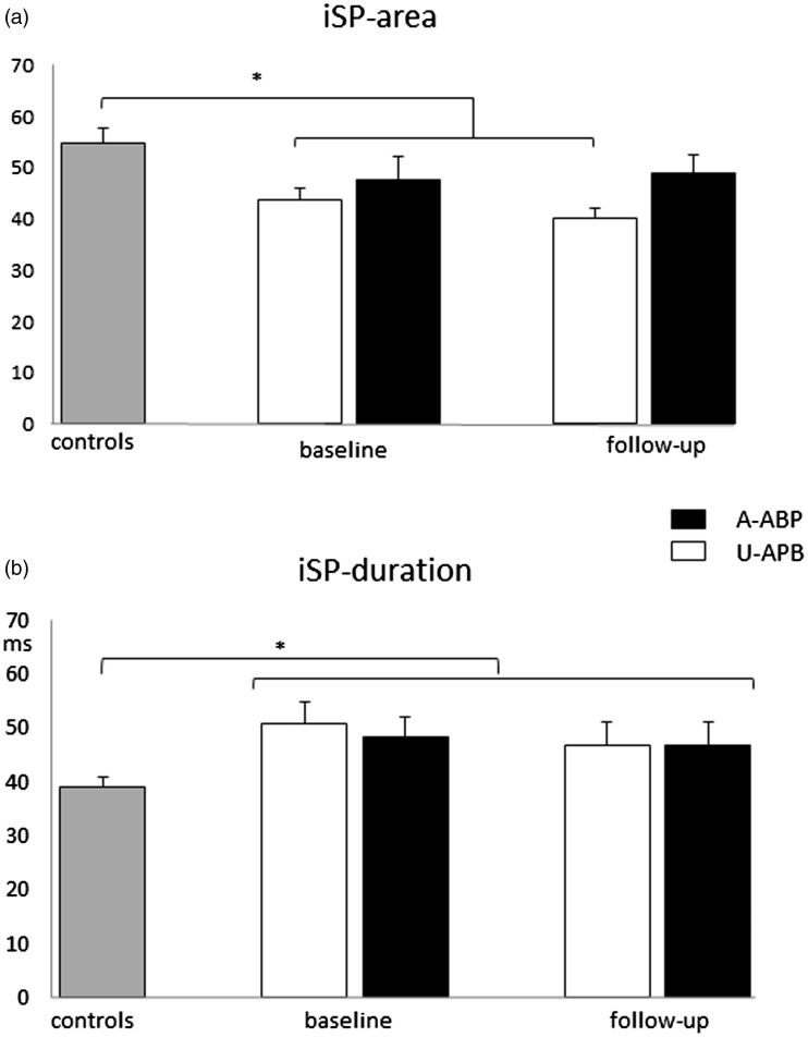
Methods: Twenty healthy and 13 relapsing-remitting MS subjects with a first motor relapse were included. TMS mapping and ipsilateral silent period (iSP) were performed after relapse and at 6-month follow-up.

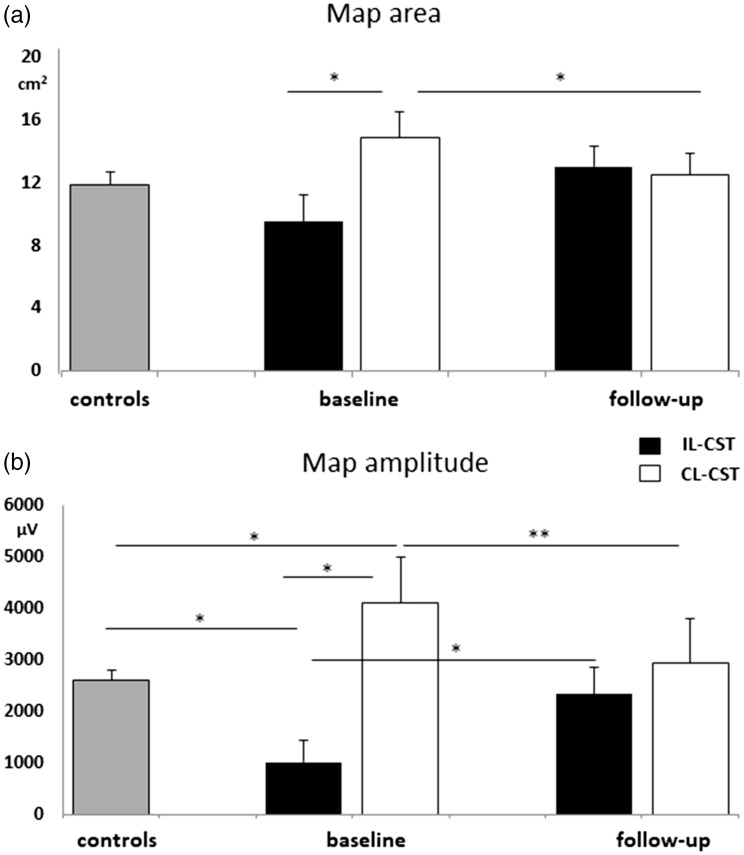
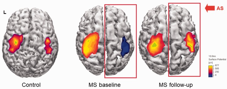
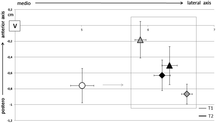
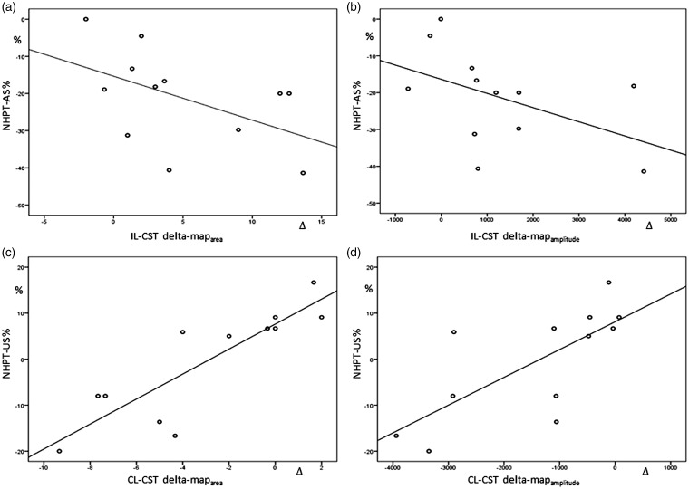
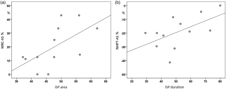
Results: Strength and dexterity of the paretic hand were impaired at baseline and improved over time. After relapse, mapamplitude and mapdensity were decreased for the ipsilesional-corticospinal tract (IL-CST) while expanded for the contralesional-CST (CL-CST). At follow-up, map parameters normalized for the CL-CST independently from recovery while the increase of outputs from the IL-CST was associated with straight and dexterity improvement. iSP measurements were impaired in MS irrespective of the phase of the disease. Prolonged iSPduration at baseline was associated with less dexterity recovery.

Conclusions: After a motor relapse, TMS mapping shows acute changes in corticospinal excitability and rearrangements of motor outputs. iSP is less influenced by the phase of disease but may better predict recovery, possibly reflecting the integrity of interhemispheric motor networks.

Keywords: TMS; iSP; mapping; multiple sclerosis; relapse.


Figures

Figure 1.
Figure 1.
Cortical map parameters in MS patients and controls. (a) greater maparea to the contralesional-corticospinal tract (CL-CST) in comparison with the ipsilesional-corticospinal tract (IL-CST) at baseline that significantly decrease at follow-up. (b) reduced mapamplitude to the IL-CST in comparison with the CL-CST and controls and increased mapamplitude to the CL-CST in comparison with controls at baseline. Significant mapamplitude increase for the IL-CST and decrease for the CL-CST at follow-up. *p <0.05; **p <0.005
Figure 2.
Figure 2.
Example of cortical motor mapping from a single patient. MEPs were obtained from the APB muscle of both sides by TMS of the contralateral motor cortex. MEPs amplitudes higher than 50 µV were interpolated and projected on an average brain cortical surface reconstruction using Curry software V4.6.
Figure 3.
Figure 3.
CoGs position in respect with vertex for controls (circular marker) and MS subjects (triangle for the unaffected side and rhombus for the affected side) at baseline (T1) and follow-up (T2). CoGs in MS subjects were laterally displaced in comparison with controls (p <0.0001). At baseline, CoG of the unaffected side was more medially (p <0.0001) and anteriorly (p =0.03) positioned than in the affected side in MS. At follow-up CoGs positions of the two hemispheres were more symmetric.
Figure 4.
Figure 4.
Ipsilateral silent period parameters (area-a and duration-b) in controls and MS on the affected (A-APB) and unaffected APB (U-APB). *p <0.05.
Figure 5.
Figure 5.
NHPT improvement for the affected side (AS) correlated with the over-time increase of maparea (a) and mapamplitude (b) to the IL-CST (r = −0.6, p =0.037 and r = −0.5, p =0.044, respectively). Amelioration in performing NHPT with the unlesioned side (US) correlated with reduction of maparea (c) and mapamplitude (d) to the CL-CST (r =0.9, p <0.001 and r =0.8, p =0.003, respectively).
Figure 6.
Figure 6.
(a) iSParea on the affected APB at baseline directly correlated with muscular straight improvement of the affected side (AS) (r =0.7, p =0.015) and (b) iSPduration with NHPT improvement of the AS at follow-up (r =0.6, p =0.035).

References

    1. Smith KJ, McDonald WI. The pathophysiology of multiple sclerosis: The mechanisms underlying the production of symptoms and the natural history of the disease. Philos Trans R Soc Lond B Biol Sci 1999; 354(1390): 1649–1673. - PMC - PubMed
    1. Filippi M, Grossman RI. MRI techniques to monitor MS evolution: The present and the future. Neurology 2002; 58(8): 1147–1153. - PubMed
    1. Mezzapesa DM, Rocca MA, Rodegher M, et al. Functional cortical changes of the sensorimotor network are associated with clinical recovery in multiple sclerosis. Hum Brain Mapp 2008; 29(5): 562–573. - PMC - PubMed
    1. Filippi M, Preziosa P, Rocca MA. Brain mapping in multiple sclerosis: Lessons learned about the human brain. Neuroimage 2017; 190: 32--2. - PubMed
    1. Chieffo R, Inuggi A, Straffi L, et al. Mapping early changes of cortical motor output after subcortical stroke: A transcranial magnetic stimulation study. Brain Stimul 2013; 6(3): 322–329. - PubMed

LinkOut - more resources