. 2020 Feb;21(1):29-43.
doi: 10.1007/s10522-019-09841-2. Epub 2019 Oct 9.

IL-6 deficiency attenuates p53 protein accumulation in aged male mouse hippocampus

Affiliations

IL-6 deficiency attenuates p53 protein accumulation in aged male mouse hippocampus

Izabela Bialuk et al. Biogerontology. 2020 Feb.

Abstract

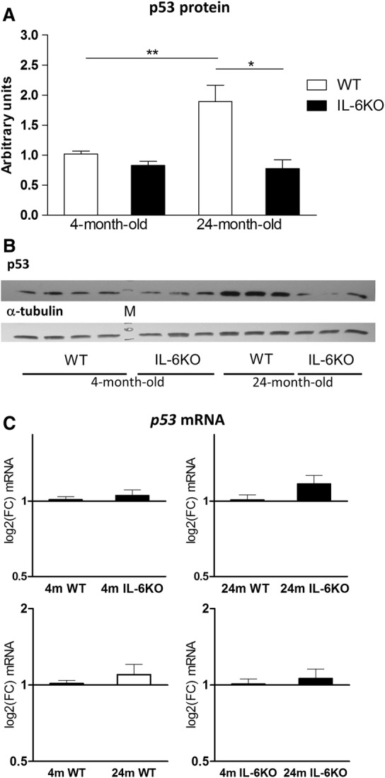
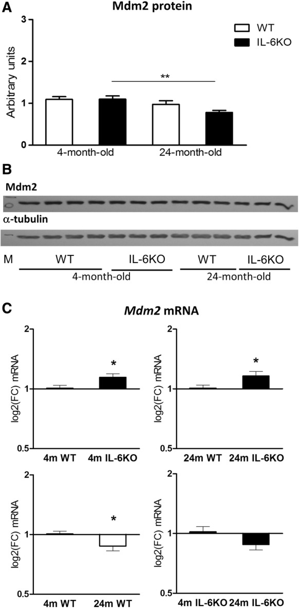
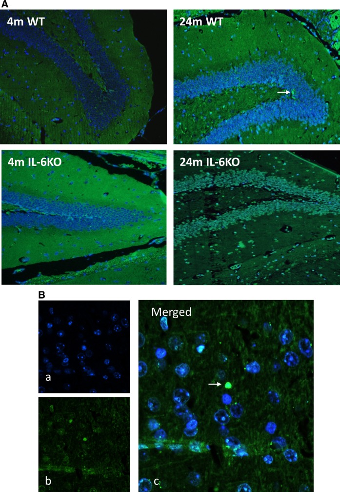
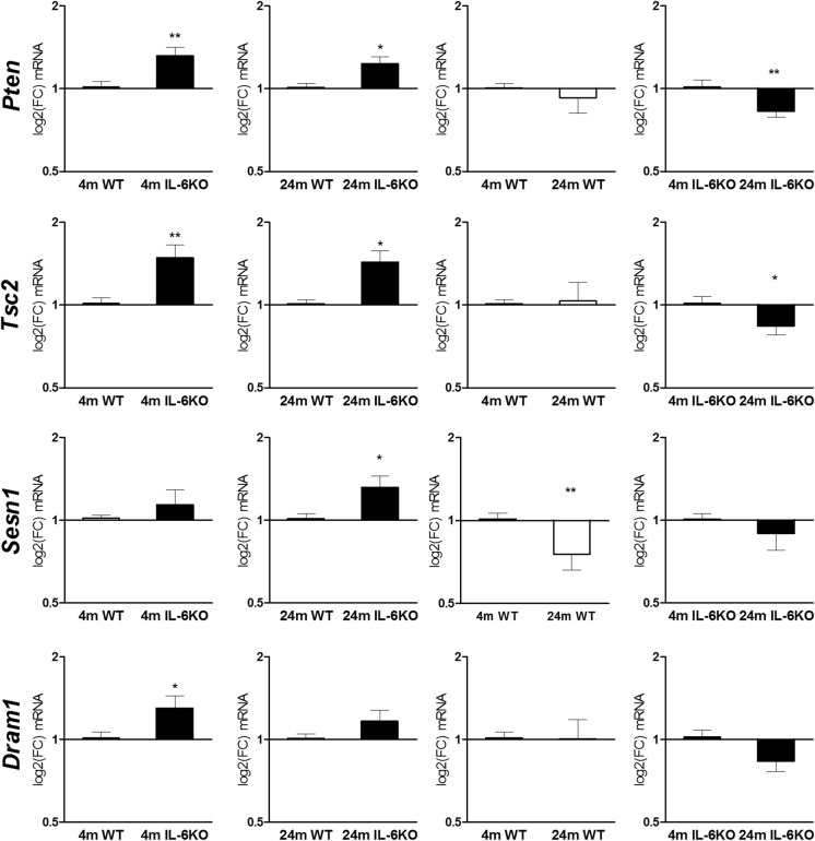
Our earlier studies demonstrated slower age-related memory decline in IL-6-deficient than in control mice. Therefore, in the present study we evaluated the effect of IL-6 deficiency and aging on expression of p53, connected with accumulation of age-related cellular damages, in hippocampus of 4- and 24-month-old IL-6-deficient C57BL/6J (IL-6KO) and wild type control (WT) mice. The accumulation of p53 protein in hippocampus of aged IL-6KO mice was significantly lower than in aged WT ones, while p53 mRNA level was significantly higher in IL-6-deficient mice, what indicates that the effect was independent on p53 transcription. Presence of few apoptotic cells in hippocampal dentate gyrus and lack of changes in levels of pro-apoptotic Bax, antiapoptotic Bcl-2, as well as in p21 protein in aged animals of both genotypes, points to low transcriptional activity of p53, especially in aged WT mice. Because the amount of p53 protein did not correlate with the level of Mdm2 protein, its main negative regulator, other than Mdm2-dependent mechanism was involved in p53 build-up. Significantly higher mRNA levels of autophagy-associated genes: Pten, Tsc2, and Dram1 in IL-6KO mice, in conjunction with significantly lower amount of Bcl-2 protein in 4-month-old IL-6KO mice, suggests that lack of IL-6/STAT3/Bcl-2 signaling could account for better autophagy performance in these mice, preventing excessive accumulation of proteins. Taken together, attenuated p53 protein build-up, absence of enhanced apoptosis, and transcriptional up-regulation of autophagy-associated genes imply that IL-6 deficiency may protect hippocampus from age-related accumulation of cellular damages.

Keywords: Apoptosis; Autophagy; Hippocampus; IL-6 deficiency; p53.


Conflict of interest statement

The authors declare that they have no conflict of interest.

Figures

Fig. 1
Fig. 1
Amount of p53 protein (a) and its mRNA transcripts (c) in hippocampus of 4- and 24-month-old IL-6-deficient (IL-6KO) and wild type control (WT) mice. Level of mRNA expression was defined as log2(FC), where FC stands for fold-change difference in mRNA level between indicated groups. Bars represent mean ± SEM obtained from six animals in each group. Expression of p53 protein was low and comparable in 4-month-old groups of both genotypes. In 24-month-old WT mice the amount of p53 protein increased significantly in comparison with 4-month-old WT (**p < 0.01) and 24-month-old IL-6KO (*p < 0.05) animals (Kruskal–Wallis with Dunn’s post hoc test). According to GLM the accumulation of p53 protein was both genotype- and age-dependent (p < 0.005 and p < 0.05, respectively). c There were no significant differences in the p53 mRNA levels between four groups of mice, however, according to GLM analysis the p53 mRNA level turned out to be influenced by genotype (p < 0.05). b Representative immunoblot for p53 protein is shown together with α-tubulin as a loading control. M molecular weight marker
Fig. 2
Fig. 2
Amount of Mdm2 protein (a) and its mRNA transcripts (c) in hippocampus of 4- and 24-month-old IL-6-deficient (IL-6KO) and wild type control (WT) mice. Level of mRNA expression was defined as log2(FC), where FC stands for fold-change difference in mRNA level between indicated groups. Bars represent mean ± SEM obtained from six animals in each group. There were no significant differences in the Mdm2 protein level in young adult mice, and aging was associated with decrease in its amount, which was statistically significant in IL-6-deficient mice in comparison with age-matched WT controls (**p < 0.01, ANOVA with Bonferroni post hoc). GLM analysis revealed significant influence of age on the Mdm2 protein abundance (p < 0.001). (c) In both 4- and 24-month-old IL-6-deficient mice Mdm2 mRNA level was significantly higher in comparison with respective control WT animals (*p < 0.05), and aging was associated with significant decrease in Mdm2 mRNA in WT controls (*p < 0.05, Wilcoxon signed rank test). According to GLM analysis the expression of Mdm2 mRNA was both genotype- and age-dependent (p < 0.005 and p < 0.001, respectively). b Representative immunoblot for Mdm2 protein is shown together with α-tubulin as a loading control. M molecular weight marker
Fig. 3
Fig. 3
a Representative microphotographs of Hoechst 33258 and TUNEL staining of hippocampal dentate gyrus form 4- and 24-month-old IL-6-deficient (IL-6KO) and wild type control (WT) mice revealed single apoptotic cells in hippocampus of aged mice of both genotypes, (magnification × 200). b (a) Fluorescent Hoechst 33258 (blue) nuclear staining, (b) TUNEL staining (green) indicating apoptotic nucleus, (c) Merged images of Hoechst 33258 (blue) and TUNEL (green) staining, (magnification × 400). White arrow indicates apoptotic cell
Fig. 4
Fig. 4
a Amount of glial fibrillary acidic protein (GFAP) in hippocampus of 4- and 24-month-old IL-6-deficient (IL-6KO) and wild type control (WT) mice was comparable in both young adult groups and insignificantly higher in both aged groups. Bars represent mean ± SEM obtained from six animals in each group. b Representative immunoblot for GFAP protein is shown together with α-tubulin as a loading control. M molecular weight marker. c Tissue staining of GFAP, an astrocytic marker (green), showed similar intensity in both 4-month-old groups and its moderate increase in both 24-month-old groups (magnification, × 20), dIL-6 mRNA in hippocampus was significantly higher in 24-month-old WT mice in comparison with 4-month-old WT ones (*p < 0.05, Wilcoxon signed rank test). Level of mRNA expression was defined as log2(FC), where FC stands for fold-change difference in mRNA level
Fig. 5
Fig. 5
mRNA transcript levels of Pten, Tsc2, Sesn1 and Dram1 genes in hippocampus of 4- and 24-month-old IL-6-deficient (IL-6KO) and wild type control (WT) mice. Level of mRNA expression was defined as log2(FC), where FC stands for fold-change difference in mRNA level between indicated groups. Bars represent mean ± SEM obtained from six animals in each group. Levels of Pten mRNA and Tsc2 mRNA were significantly higher in both 4- and 24-month-old IL-6KO mice in comparison with age-matched WT controls (**p < 0.01, and *p < 0.05, respectively). Aging was associated with significant decrease in Pten mRNA and Tsc2 mRNA levels (**p < 0.01, and *p < 0.05, respectively) in IL-6KO animals (Wilcoxon signed rank test). IL-6 deficiency was associated with statistically significant increase in Sesn1 mRNA in aged animals in comparison with respective WT group (*p < 0.05), while aging was associated with significantly decreased its mRNA transcript in WT control mice (**p < 0.01, Wilcoxon signed rank test). Deficiency of IL-6 resulted in significantly higher expression of Dram1 mRNA in 4-month-old mice (*p < 0.05), while aging diminished Dram1 mRNA level, but the effect was insignificant (Wilcoxon signed rank test). GLM analysis revealed influence of genotype on the transcription of Pten, Tsc2, and Dram1 (p < 0.0005, p <0.0005 and p <0.05, respectively), and influence of age on the transcription of Pten and Dram1 (p <0.01 and p <0.005, respectively)

References

    1. Aniszewska A, Chlodzinska N, Bartkowska K, Winnicka MM, Turlejski K, Djavadian RL. The expression of interleukin-6 and its receptor in various brain regions and their roles in exploratory behavior and stress responses. J Neuroimmunol. 2015;284:1–9. - PubMed
    1. Bermejo P, Martin-Aragon S, Benedi J, Susin C, Felici E, Gil P, Ribera JM, Villar AM. Differences of peripheral inflammatory markers between mild cognitive impairment and Alzheimer’s disease. Immunol Lett. 2008;117:198–202. - PubMed
    1. Best BP. Nuclear DNA damage as a direct cause of aging. Rejuvenation Res. 2009;12:199–208. - PubMed
    1. Bialuk I, Winnicka MM. Facilitatory Effect of IL-6 deficiency on long-term spatial memory in young adult mice. Behav Genet. 2018;48:236–246. - PubMed
    1. Bialuk I, Taranta A, Winnicka MM. IL-6 deficiency alters spatial memory in 4- and 24-month-old mice. Neurobiol Learn Mem. 2018;155:21–29. - PubMed

Publication types