. 2019 Oct;47(7):865-886.
doi: 10.1177/0192623319873871. Epub 2019 Oct 10.

Histology Atlas of the Developing Mouse Urinary System With Emphasis on Prenatal Days E10.5-E18.5

Affiliations

Histology Atlas of the Developing Mouse Urinary System With Emphasis on Prenatal Days E10.5-E18.5

Susan A Elmore et al. Toxicol Pathol. 2019 Oct.

Abstract

Congenital abnormalities of the urinary tract are some of the most common human developmental abnormalities. Several genetically engineered mouse models have been developed to mimic these abnormalities and aim to better understand the molecular mechanisms of disease. This atlas has been developed as an aid to pathologists and other biomedical scientists for identification of abnormalities in the developing murine urinary tract by cataloguing normal structures at each stage of development. Hematoxylin and eosin- and immunohistochemical-stained sections are provided, with a focus on E10.5-E18.5, as well as a brief discussion of postnatal events in urinary tract development. A section on abnormalities in the development of the urinary tract is also provided, and molecular mechanisms are presented as supplementary material. Additionally, overviews of the 2 key processes of kidney development, branching morphogenesis and nephrogenesis, are provided to aid in the understanding of the complex organogenesis of the kidney. One of the key findings of this atlas is the histological identification of the ureteric bud at E10.5, as previous literature has provided conflicting reports on the initial point of budding. Furthermore, attention is paid to points where murine development is significantly distinct from human development, namely, in the cessation of nephrogenesis.

Keywords: atlas; bladder; development; embryo; genetically engineered mice; kidney; urethra.


Conflict of interest statement

Declaration of Conflicting Interests Statement

The author(s) declared no potential, real, or perceived conflicts of interest with respect to the research, authorship, and/or publication of this article.

Figures

Figure 1
Figure 1
Schematic representation of early kidney development in the mouse embryo. The kidney develops as three portions in a cranio-caudal distribution from the intermediate mesoderm following gastrulation in mammals: the pronephros, mesonephros, and metanephros. The pronephros and mesonephros are transient structures. The pronephros sprouts from the anterior Wolffian duct at around E8.0 and degenerates by E9.0, when the mesonephros begins to develop. The mesonephros consists of the Wolffian duct and a linear array of mesonephric mesenchyme, with only the cranial portion developing into mesonephric tubules. The mesonephros begin to degenerate at around E14.5 and quickly disappears via apoptosis in a caudal to cranial direction. The metanephric mesenchyme arises around E10.5 and the metanephros persists as the definitive adult kidney.
Figure 2
Figure 2
Representative sagittal and transverse sections of an E10.5 embryo during metanephric development. Sagittal sections highlight the ureteric bud (UB) and metanephric mesenchyme (MM) at low (2A) and high (2B) magnifications. Transverse sections depict mesonephric tubules (MST) at low (C) and high (D) magnifications. MS = mesonephros.
Figure 3
Figure 3
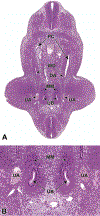
Representative transverse sections of an E11.5 embryo. Sections show bifurcation of the ureteric bud (UB) at low (3A) and high (3B) magnifications. DA = dorsal aorta, WD = Wolffian (mesonephric) duct, PC = peritoneal cavity, MM = metanephric mesenchyme, UA = umbilical aorta.
Figure 4
Figure 4
Diagrammatic representation of nephrogenesis illustrating the configurational changes as nephrons develop. Reprinted with permission from the Journal of Toxicologic Pathology.
Figure 5
Figure 5
Representative sagittal and transverse sections of an E12.5 embryo. Sections show early branching morphogenesis (5A), early renal vesicles (5B) and the connecting of the common nephric duct to the urogenital sinus (5C and D). Figure 5A sagittal section shows that the ureteric bud has expanded within the metanephric mesenchyme to form the primitive renal pelvis, and bifurcation (asterisks) continues to form the future intrarenal collecting system. A new ampulla (5A) can be seen surrounded by MM. In Figure 5B, renal vesicles (RV), precursors to glomeruli, can be seen in this transverse section. Figure 5C sagittal section depicts the common nephric duct (CND) connecting with the urogenital sinus (UGS). Ureter maturation depends on apoptotic elimination of the CND via apoptosis. Figure 5D, a higher magnification of C, shows that the CND connects with, but does not yet communicate with, the UGS. GT = genital tubercle, HG = hindgut, UM = umbilicus.
Figure 6
Figure 6
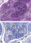
Representative sagittal and frontal sections from an E13.5 embryo. Figure 6A is a sagittal section that shows developing renal vesicles (RV) and metanephric tubules (MTT) within a more distinct cortical region. In Figure 6B, (frontal section) macrophages (arrows), highlighted using an F480 immunohistochemical stain, can be seen interspersed within the interstitial tissue of the metanephros. UR = ureter.
Figure 7
Figure 7
Representative sagittal section of the developing kidney at E14.5. Section shows primitive glomeruli (PG) in the outer cortex, collecting ducts (CD), and early stromal cells in the medulla (MD).
Figure 8
Figure 8
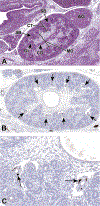
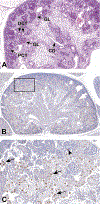
Representative sagittal sections of the developing kidney at E15.5. Figure 8A shows the medulla (MD), which has become more distinct at this stage as it becomes devoid of glomeruli. As the glomeruli continue to develop, a variety of shapes can be appreciated including S shaped bodies (SB; arrows) and more mature glomeruli (GL). In Figure 8B, Class III β-tubulin antibody shows strong immunoreactivity in perivascular cells (arrows) in a kidney from an E15.5 embryo. Figure 8C shows a higher magnification illustrating cytoplasmic localization in specific pericytes (arrows). CD = collecting duct, AG = adrenal gland, CT = cortex.
Figure 9
Figure 9
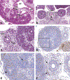
Representative transverse and frontal sections of the developing kidney at E16.5. Primitive glomeruli (PG) in the cortex (CT), collecting ducts (CD) in the medulla, renal pelvis (RP) and the connecting ureter (UR) are shown in a transverse section (9A). A lower magnification frontal section (9B) shows the left and right kidneys (K) and the connecting ureters (UR) on either side of the abdominal aorta (AA). A higher magnification frontal section (9C) shows the multilayered transitional urothelium (URO) within the ureter. The mesenchymal layer (ML) will further differentiate into stromal cells, smooth muscle cells, and adventitial fibroblasts. In frontal sections Figures 9D–F, anti-nestin antibody reveals strong immunoreactivity in the vascular cleft of the developing S-shaped glomeruli, and in the condensing mesenchyme surrounding the ureter at E16.5. Figure 9E is a higher magnification of the boxed region in 9D showing cytoplasmic staining in the vascular cleft of an S-shaped glomerulus (arrow) and in the cortical mesenchyme surrounding ureteric branches (arrowhead). Figure 9F shows cytoplasmic staining in the mesenchymal tissue surrounding a ureter (arrows).
Figure 10.
Figure 10.
Representative sagittal and frontal sections of the developing kidney at E18.5. By E18.0, the kidneys are in an advanced stage of differentiation, with increased numbers of more mature glomeruli and larger numbers of proximal and distal convoluted tubules within the renal cortex. Figure 10A is a sagittal section of the developing kidney at E18.5 and shows the collecting duct (CD) and, within the cortex, more mature glomeruli (GL), proximal convoluted tubules (PCT) with brush borders and distal convoluted tubules (DCT). Figure 10B is a frontal section stained with Ki67. Figure10C is a higher magnification of boxed area in 10B and shows a subset of renal tubular cells and occasional glomeruli that are positive for Ki67 at E18.5. Arrows point to positive nuclei in tubular epithelial cells. Arrowhead shows one positive nucleus within a glomerulus.
Figure 11
Figure 11
Representative transverse section of the developing lower urinary tract at E10.5. This section shows the cloaca (C), dorsal aorta (DA), coelomic cavity (CC) and mesonephric tubules (arrows). The most caudal region of the hindgut begins to dilate between E8.5 and E9.0, forming a region that is significantly expanded when compared to the midgut and cranial portions of the hindgut. By E10.5, this expansion can be definitively identified as the cloaca. The urorectal septum has not yet divided the cloaca into the urogenital sinus and anorectal canal.
Figure 12
Figure 12
Representative sagittal section of the developing lower urinary tract at E11.5. The ventral portion of the cloaca, which is destined to become the urogenital sinus, becomes more distinct from the hindgut by the continued down-growth of the urorectal septum and the genital tubercle is visible as a protrusion adjacent to the cloaca. This sagittal section shows the undivided cloaca (C), urorectal septum (URS), hindgut (HG; anorectal canal) and the now-visible genital tubercle (GT).
Figure 13
Figure 13
Representative sagittal section of the developing lower urinary tract at E12.5. The cloaca is not fully divided until E13.5. A portion of the ventral urogenital sinus (UGS) region of the cloaca extends toward the umbilicus (UM) to form a tubular structure called the urachus (not shown in section). This section shows the transposition of the common nephric duct (CND) to the future bladder wall and the urachus. GT = genital tubercle, HG = hindgut (anorectal canal).
Figure 14
Figure 14
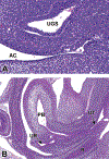
Representative sagittal sections of the developing lower urinary tract at E14.5. Figure 14A shows that the cloacal membrane disintegrated by apoptosis one day earlier at E13.5, completing the division of the cloaca into the urogenital sinus (UGS) and anorectal canal (AC). Figure 14B shows that at E14.5 the vesicourethral canal continues to develop into the primitive bladder (PB) and the pelvic portion of the urethra (UT). The ureter (UR) has also opened completely to the primitive bladder. R = rectum.
Figure 15
Figure 15
Representative sagittal and frontal images of the developing lower urinary tract at E15.5. The sagittal section (15A) shows the mature bladder (BL), the ureter (UR) in the region of the neck of the bladder, the rectum (R) and genital tubercle (GT). The frontal section (15B) shows that the urinary bladder has reached a mature stage by E15.5, with a multilayered transitional urothelium (URO), lamina propria (LP), and smooth muscle (SM). AC = anorectal canal.
Figure 16
Figure 16
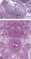
Representative frontal section of the male developing lower urinary tract at E16.5. The proximal urethral meatus (PUM) continues to close during E16.5. Fusion of the urethral folds (UF) in the ventral midline leads to the formation of the urethral groove and subsequently the penile portion of male urethra (UT). GT = genital tubercle.
Figure 17
Figure 17
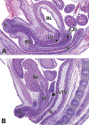
Representative sagittal sections of the male (17A) and female (17B) developing lower urinary tract at E18.5. Both figures show the urethra (UT) connecting to the bladder (BL). Other structures such as the rectum (R), ejaculatory duct (EJD), penis (P), uterus (UTS), bulbourethral gland (BG) and clitoris (CL) are labeled for orientation.

Similar articles

Cited by

References

    1. Pohl M, Bhatnagar V, Mendoza SA, Nigam SK. Toward an etiological classification of developmental disorders of the kidney and upper urinary tract. Kidney Int 2002;61(1):10–19. - PubMed
    1. Michos O. Kidney development: from ureteric bud formation to branching morphogenesis. Curr Opin Genet Dev 2009;19(5):484–490. - PMC - PubMed
    1. North American Pediatric Renal Trials and Collaborative Studies. 2008 Annual Report 2008.
    1. Rasouly HM, Lu W. Lower urinary tract development and disease. Wiley Interdiscip Rev Syst Biol Med 2013;5(3):307–342. - PMC - PubMed
    1. Sanna-Cherchi S, Caridi G, Weng PL, et al. Genetic approaches to human renal agenesis/hypoplasia and dysplasia. Pediatr Nephrol 2007;22(10):1675–1684. - PMC - PubMed

Publication types

LinkOut - more resources