

Robot controlled, continuous passive movement of the ankle reduces spinal cord excitability in participants with spasticity: a pilot study
- PMID: 31599345
- PMCID: PMC6882765
- DOI: 10.1007/s00221-019-05662-4
Robot controlled, continuous passive movement of the ankle reduces spinal cord excitability in participants with spasticity: a pilot study
Abstract
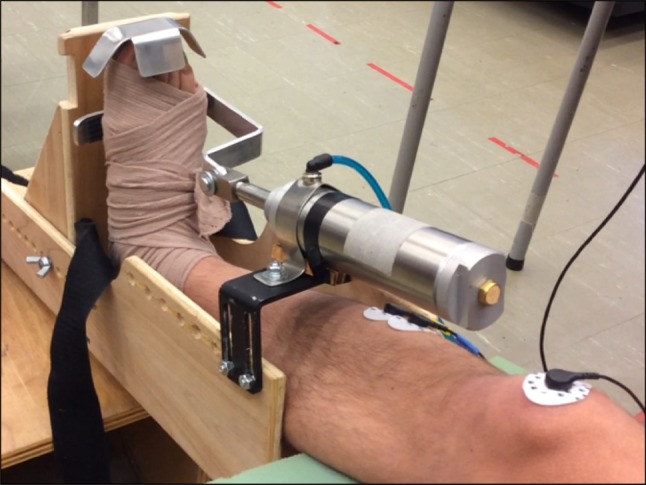
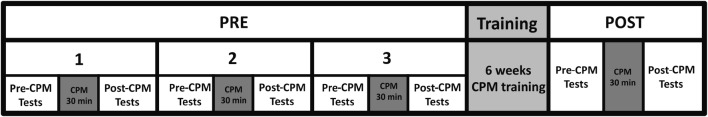
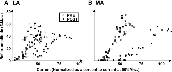
Spasticity of the ankle reduces quality of life by impeding walking and other activities of daily living. Robot-driven continuous passive movement (CPM) is a strategy for lower limb spasticity management but effects on spasticity, walking ability and spinal cord excitability (SCE) are unknown. The objectives of this experiment were to evaluate (1) acute changes in SCE induced by 30 min of CPM at the ankle joint, in individuals without neurological impairment and those with lower limb spasticity; and, (2) the effects of 6 weeks of CPM training on SCE, spasticity and walking ability in those with lower limb spasticity. SCE was assessed using soleus Hoffmann (H-) reflexes, collected prior to and immediately after CPM for acute assessments, whereas a multiple baseline repeated measures design assessed changes following 18 CPM sessions. Spasticity and walking ability were assessed using the Modified Ashworth Scale, the 10 m Walk test, and the Timed Up and Go test. Twenty-one neurologically intact and nine participants with spasticity (various neurological conditions) were recruited. In the neurologically intact group, CPM caused bi-directional modulation of H-reflexes creating 'facilitation' and 'suppression' groups. In contrast, amongst participants with spasticity, acute CPM facilitated H-reflexes. After CPM training, H-reflex excitability on both the more-affected and less-affected sides was reduced; on the more affected side H@Thres, H@50 and H@100 all significantly decreased following CPM training by 96.5 ± 7.7%, 90.9 ± 9.2%, and 62.9 ± 21.1%, respectively. After training there were modest improvements in walking and clinical measures of spasticity for some participants. We conclude that CPM of the ankle can significantly alter SCE. The use of CPM in those with spasticity can provide a temporary period of improved walking, but efficacy of treatment remains unknown.
Keywords: Continuous passive movement; H-reflex; Spasticity; Spinal cord excitability.
Conflict of interest statement
The authors have no conflicts of interest to declare, financial or otherwise.
Figures


References
-
- Alonso RJ, Mancall EL. The clinical management of spasticity. Semin Neurol. 1991;11:215–219. - PubMed
-
- Barnes CD, Pompeiano O. Effects of muscle vibration on the pre- and postsynaptic components of the extensor monosynaptic reflex. Brain Res. 1970;18:384–388. - PubMed
-
- Bohannon RW, Smith MB. Interrater reliability of a modified Ashworth scale of muscle spasticity. Phys Ther. 1987;67:206–207. - PubMed
-
- Bourbonnais D, Vanden Noven S. Weakness in patients with hemiparesis. Am J Occup Ther. 1989;43:313–319. - PubMed
-
- Bovend’Eerdt TJ, Newman M, Barker K, Dawes H, Minelli C, Wade DT. The effects of stretching in spasticity: a systematic review. Arch Phys Med Rehabil. 2008;89:1395–1406. - PubMed
MeSH terms
LinkOut - more resources
Full Text Sources
