

Altered inter- and intrahemispheric functional connectivity dynamics in autistic children
- PMID: 31600014
- PMCID: PMC7268059
- DOI: 10.1002/hbm.24812
Altered inter- and intrahemispheric functional connectivity dynamics in autistic children
Abstract
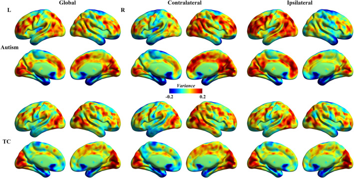
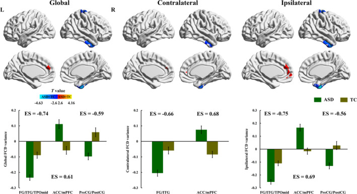
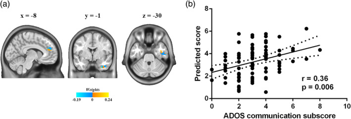
Emerging evidence has associated autism spectrum disorder (ASD) with static functional connectivity abnormalities between multiple brain regions. However, the temporal dynamics of intra- and interhemispheric functional connectivity patterns remain unknown in ASD. Resting-state functional magnetic resonance imaging data were analyzed for 105 ASD and 102 demographically matched typically developing control (TC) children (age range: 7-12 years) available from the Autism Brain Imaging Data Exchange database. Whole-brain functional connectivity was decomposed into ipsilateral and contralateral functional connectivity, and sliding-window analysis was utilized to capture the intra- and interhemispheric dynamic functional connectivity density (dFCD) patterns. The temporal variability of the functional connectivity dynamics was further quantified using the standard deviation (SD) of intra- and interhemispheric dFCD across time. Finally, a support vector regression model was constructed to assess the relationship between abnormal dFCD variance and autism symptom severity. Both intra- and interhemispheric comparisons showed increased dFCD variability in the anterior cingulate cortex/medial prefrontal cortex and decreased variability in the fusiform gyrus/inferior temporal gyrus in autistic children compared with TC children. Autistic children additionally showed lower intrahemispheric dFCD variability in sensorimotor regions including the precentral/postcentral gyrus. Moreover, aberrant temporal variability of the contralateral dFCD predicted the severity of social communication impairments in autistic children. These findings demonstrate altered temporal dynamics of the intra- and interhemispheric functional connectivity in brain regions incorporating social brain network of ASD, and highlight the potential role of abnormal interhemispheric communication dynamics in neural substrates underlying impaired social processing in ASD.
Keywords: autism spectrum disorder; dynamic functional connectivity; interhemisphere; intrahemisphere; resting-state functional magnetic resonance imaging.
© 2019 The Authors. Human Brain Mapping published by Wiley Periodicals, Inc.
Conflict of interest statement
None declared.
Figures
Similar articles
-
Abnormal interhemispheric and intrahemispheric functional connectivity dynamics in drug-naïve first-episode schizophrenia patients with auditory verbal hallucinations.Hum Brain Mapp. 2022 Oct 1;43(14):4347-4358. doi: 10.1002/hbm.25958. Epub 2022 May 25. Hum Brain Mapp. 2022. PMID: 35611547 Free PMC article.
-
More Than Just Statics: Temporal Dynamic Changes in Inter- and Intrahemispheric Functional Connectivity in First-Episode, Drug-Naive Patients With Major Depressive Disorder.Front Hum Neurosci. 2022 Apr 8;16:868135. doi: 10.3389/fnhum.2022.868135. eCollection 2022. Front Hum Neurosci. 2022. PMID: 35463932 Free PMC article.
-
Alternations voxel-wise interhemispheric and intrahemipheric functional connectivity dynamics in internet gaming disorder.J Affect Disord. 2025 Jan 15;369:662-670. doi: 10.1016/j.jad.2024.10.055. Epub 2024 Oct 16. J Affect Disord. 2025. PMID: 39419186
-
Resting-state abnormalities of posterior cingulate in autism spectrum disorder.Prog Mol Biol Transl Sci. 2020;173:139-159. doi: 10.1016/bs.pmbts.2020.04.010. Epub 2020 May 4. Prog Mol Biol Transl Sci. 2020. PMID: 32711808 Review.
-
Brain Connectivity and Neuroimaging of Social Networks in Autism.Trends Cogn Sci. 2018 Dec;22(12):1103-1116. doi: 10.1016/j.tics.2018.09.008. Epub 2018 Oct 31. Trends Cogn Sci. 2018. PMID: 30391214 Free PMC article. Review.
Cited by
-
Reduced Inter-hemispheric Resting State Functional Connectivity and Its Association With Social Deficits in Autism.Front Psychiatry. 2021 Mar 4;12:629870. doi: 10.3389/fpsyt.2021.629870. eCollection 2021. Front Psychiatry. 2021. PMID: 33746796 Free PMC article. Review.
-
An Investigation of Age-related Neuropathophysiology in Autism Spectrum Disorder Using Fixel-based Analysis of Corpus Callosum White Matter Micro- and Macrostructure.J Autism Dev Disord. 2024 Jun;54(6):2198-2210. doi: 10.1007/s10803-023-05980-1. Epub 2023 Apr 20. J Autism Dev Disord. 2024. PMID: 37079181 Free PMC article.
-
Dysfunction in the Interaction of Information Between and Within the Bilateral Primary Sensory Cortex.Front Aging Neurosci. 2022 Apr 8;14:862107. doi: 10.3389/fnagi.2022.862107. eCollection 2022. Front Aging Neurosci. 2022. PMID: 35462694 Free PMC article.
-
Abnormal interhemispheric and intrahemispheric functional connectivity dynamics in drug-naïve first-episode schizophrenia patients with auditory verbal hallucinations.Hum Brain Mapp. 2022 Oct 1;43(14):4347-4358. doi: 10.1002/hbm.25958. Epub 2022 May 25. Hum Brain Mapp. 2022. PMID: 35611547 Free PMC article.
-
Cofluctuation analysis reveals aberrant default mode network patterns in adolescents and youths with autism spectrum disorder.Hum Brain Mapp. 2022 Oct 15;43(15):4722-4732. doi: 10.1002/hbm.25986. Epub 2022 Jul 4. Hum Brain Mapp. 2022. PMID: 35781734 Free PMC article.
References
-
- American Psychiatric Association . (2013). Diagnostic and statistical manual of mental disorders (DSM‐5®). Arlington, VA: American Psychiatric Pub.
