Mechanical effects of MitraClip on leaflet stress and myocardial strain in functional mitral regurgitation - A finite element modeling study
- PMID: 31600276
- PMCID: PMC6786765
- DOI: 10.1371/journal.pone.0223472
Mechanical effects of MitraClip on leaflet stress and myocardial strain in functional mitral regurgitation - A finite element modeling study
Abstract
Purpose: MitraClip is the sole percutaneous device approved for functional mitral regurgitation (MR; FMR) but MR recurs in over one third of patients. As device-induced mechanical effects are a potential cause for MR recurrence, we tested the hypothesis that MitraClip increases leaflet stress and procedure-related strain in sub-valvular left ventricular (LV) myocardium in FMR associated with coronary disease (FMR-CAD).
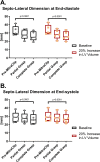
Methods: Simulations were performed using finite element models of the LV + mitral valve based on MRI of 5 sheep with FMR-CAD. Models were modified to have a 20% increase in LV volume (↑LV_VOLUME) and MitraClip was simulated with contracting beam elements (virtual sutures) placed between nodes in the center edge of the anterior (AL) and posterior (PL) mitral leaflets. Effects of MitraClip on leaflet stress in the peri-MitraClip region of AL and PL, septo-lateral annular diameter (SLAD), and procedure-related radial strain (Err) in the sub-valvular myocardium were calculated.
Results: MitraClip increased peri-MitraClip leaflet stress at end-diastole (ED) by 22.3±7.1 kPa (p<0.0001) in AL and 14.8±1.2 kPa (p<0.0001) in PL. MitraClip decreased SLAD by 6.1±2.2 mm (p<0.0001) and increased Err in the sub-valvular lateral LV myocardium at ED by 0.09±0.04 (p<0.0001)). Furthermore, MitraClip in ↑LV_VOLUME was associated with persistent effects at ED but also at end-systole where peri-MitraClip leaflet stress was increased in AL by 31.9±14.4 kPa (p = 0.0268) and in PL by 22.5±23.7 kPa (p = 0.0101).
Conclusions: MitraClip for FMR-CAD increases mitral leaflet stress and radial strain in LV sub-valvular myocardium. Mechanical effects of MitraClip are augmented by LV enlargement.
Conflict of interest statement
The authors have declared that no competing interests exist.
Figures


References
-
- Gorman RC, Gorman JH 3rd, Edmunds LH Jr. Ischemic mitral regurgitation in cardiac surgery in the adult In: Cohn LH, Edmunds LH Jr., editors. Cardiac Surgery in the adult. New York: McGraw-Hill; 2003. p. 1751–70.
-
- Hickey MS, Smith LR, Muhlbaier LH, Harrell FE Jr., Reves JG, Hinohara T, et al. Current prognosis of ischemic mitral regurgitation. Implications for future management. Circulation. 1988;78(3 Pt 2):I51–9. . - PubMed
-
- Abbott receives FDA approval for expanded indication for MitraClip™ device 2019. Available from: https://abbott.mediaroom.com/2019-03-14-Abbott-Receives-FDA-Approval-for....
-
- MitraClip Clip Delivery System: Abbott Vascular; 2016. Available from: http://www.abbottvascular.com/docs/ifu/structural_heart/eIFU_MitraClip.pdf.
Publication types
MeSH terms
Grants and funding
LinkOut - more resources
Full Text Sources
Research Materials
Miscellaneous

