. 2019 Oct 9;26(4):493-503.e6.
doi: 10.1016/j.chom.2019.09.012.

Zika Virus NS3 Mimics a Cellular 14-3-3-Binding Motif to Antagonize RIG-I- and MDA5-Mediated Innate Immunity

Affiliations

Zika Virus NS3 Mimics a Cellular 14-3-3-Binding Motif to Antagonize RIG-I- and MDA5-Mediated Innate Immunity

William Riedl et al. Cell Host Microbe. .

Abstract

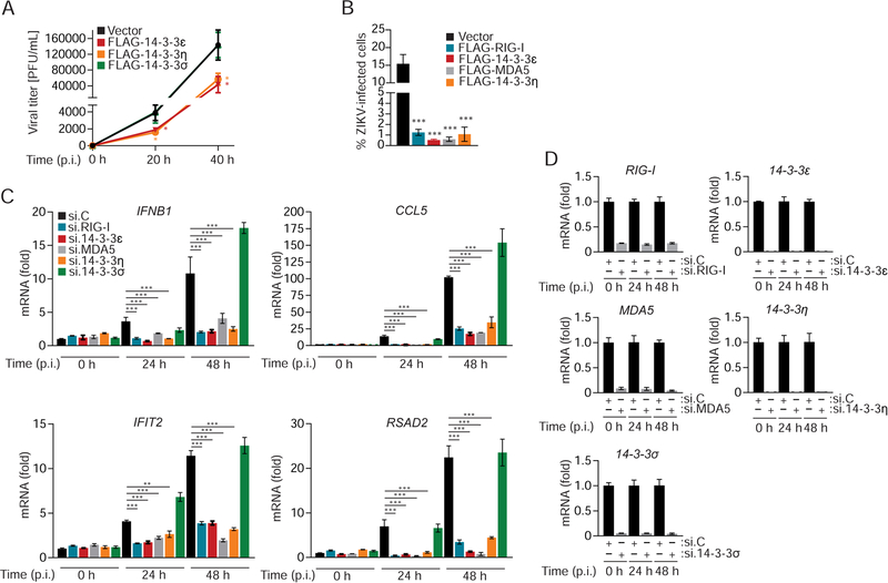
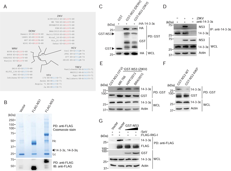
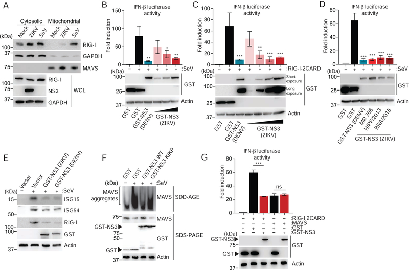
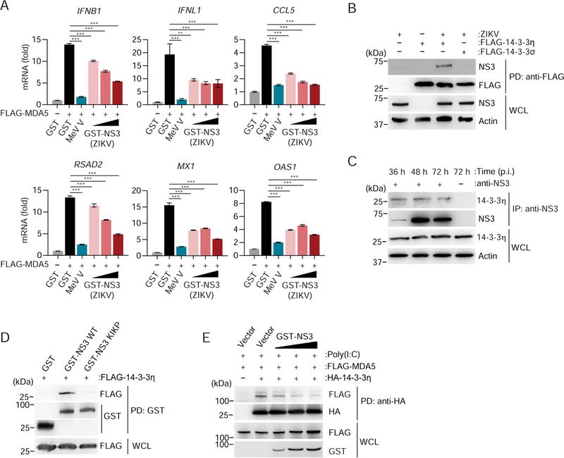
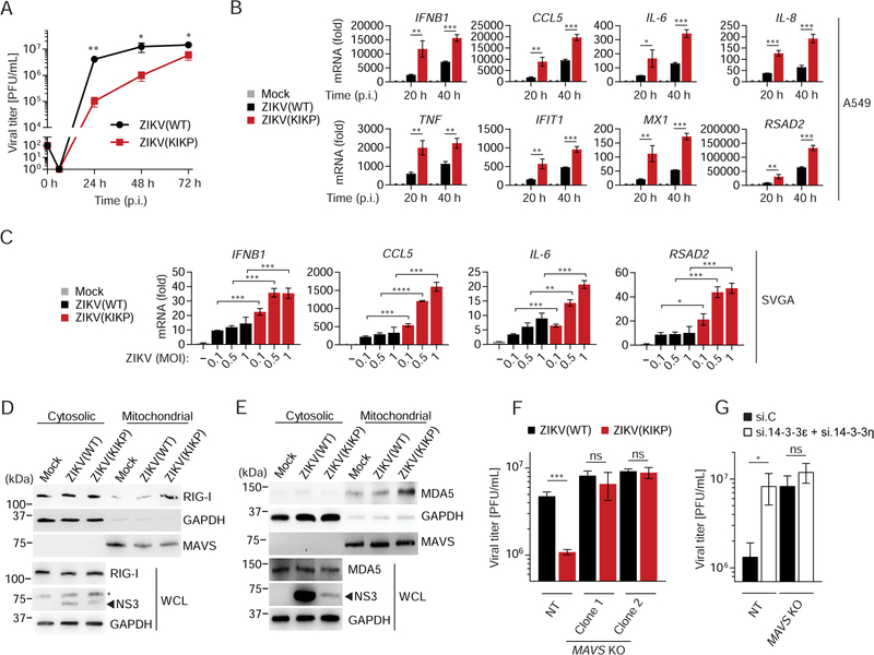
14-3-3 protein family members facilitate the translocation of RIG-I-like receptors (RLRs) to organelles that mediate downstream RLR signaling, leading to interferon production. 14-3-3ϵ promotes the cytosolic-to-mitochondrial translocation of RIG-I, while 14-3-3η facilitates MDA5 translocation to mitochondria. We show that the NS3 protein of Zika virus (ZIKV) antagonizes antiviral gene induction by RIG-I and MDA5 by binding to and sequestering the scaffold proteins 14-3-3ϵ and 14-3-3η. 14-3-3-binding is mediated by a negatively charged RLDP motif in NS3 that is conserved in ZIKV strains of African and Asian lineages and is similar to the one found in dengue and West Nile viruses. ZIKV NS3 is sufficient to inhibit the RLR-14-3-3ϵ/η interaction and to suppress antiviral signaling. Mutational perturbation of 14-3-3ϵ/η binding in a recombinant ZIKV leads to enhanced innate immune responses and impaired growth kinetics. Our study provides molecular understanding of immune evasion functions of ZIKV, which may guide vaccine and anti-flaviviral therapy development.

Keywords: RIG-I-like receptors; Zika virus; flaviviruses; innate immunity; interferon; viral immune evasion.


Conflict of interest statement

DECLARATION OF INTERESTS

M.U.G. and Y.K.C. are co-inventors on a patent application for use of the RxD/EP motif and KIKP mutant Zika virus and related viruses.

Figures

Figure 1.
Figure 1.. 14–3-3ε and 14–3-3η inhibit ZIKV replication by mediating antiviral gene expression.
(A) Viral titers in the supernatant of SVGA cells that were transfected with empty vector or the indicated FLAG-tagged 14–3-3 proteins and then infected with ZIKV (BRA/2015; MOI 0.1) for the indicated times, determined by plaque assay and presented as PFU/mL. (B) Frequency of infected A549 cells that were transfected with either empty vector or plasmids encoding the indicated FLAG-tagged proteins for 42 h and then infected with ZIKV (BRA/2015; MOI 0.2) for 48 h, assessed by flow cytometry using an antibody recognizing the ZIKV E protein. (C) qRT-PCR analysis of the indicated transcripts in SVGA cells that were transfected with the indicated siRNAs for 24 h and then infected with ZIKV (BRA/2015; MOI 1) for 24 or 48 h. (D) Silencing efficiency of indicated genes for the experiment shown in (C), determined by qRT-PCR and normalized to cellular GAPDH. Data are expressed as means ± SD (n = 3). *p < 0.05, **p < 0.01, ***p < 0.001 [Student’s t-test in (A)]; *p < 0.01, **p < 0.001, ***p < 0.0001 [ANOVA in (B, C)]. Data are representative of two (A, C, D) or three (B) independent experiments. See also Figure S1.
Figure 2.
Figure 2.. ZIKV NS3 binds to human 14–3-3ε and competes with RIG-I for the 14–3-3ε interaction.
(A) Amino acid sequence of the NS3 region containing the phosphomimetic 14–3-3-interaction motif Rx(E/D)P (residues 64–67; red) in ZIKV, DENV and WNV, as well as the corresponding NS3 regions in the related Flaviviridae family members JEV, YFV, TBEV, and HCV (blue). Representative virus strains are shown (see Figure S2A for a summary of the complete sequence analysis). Of note, cladogram shown is not to scale. (B) Upper panel: Coomassie-stained complexes of affinity-purified FLAG-tagged ZIKV NS3 (H/PF/2013) from transiently transfected HEK 293T cells using anti-FLAG agarose. Purified FLAG-tagged ZIKV NS1 (H/PF/2013) as well as cells transfected with empty vector served as controls. Asterisks denote the FLAG-NS1 and FLAG-NS3 proteins. Arrow indicates the band that identified 14–3-3ε and 14–3-3η by MS analysis. Lc, antibody light chain. Hc, antibody heavy chain. Lower panel: Anti-FLAG immunoblot (IB) analysis of affinity-purified FLAG-tagged NS3 and NS1 from the complexes shown above. Asterisks denote the FLAG-NS1 and FLAG-NS3 proteins. (C) Binding of GST-NS3 from ZIKV (H/PF/2013) and DENV (NGC; positive control) or GST (negative control) to HA-tagged 14–3-3ɛ in HEK 293T cells transiently transfected for 42 h to express those proteins, determined by GST-pulldown (PD) and IB using anti-HA and anti-GST. (D) Interaction of NS3 with endogenous 14–3-3ε in SVGA cells infected with ZIKV (BRA/2015; MOI 1) for 48 h, assessed by immunoprecipitation (IP) with anti-14–3-3ε, followed by IB with anti-NS3 and anti-14–3-3ε. Whole cell lysates (WCLs) were probed with anti-NS3, anti-14–3-3ε and anti-Actin (loading control). (E) Binding of GST-NS3 from the indicated ZIKV strains to endogenous 14–3-3ε in HEK 293T cells transfected for 42 h to express those proteins, assessed by GST-PD and IB using anti-14–3-3ε and anti-GST. Cells transfected with GST-NS3 from YFV (strain 17D) served as negative control. (F) Binding of GST-fused ZIKV NS3 (BRA/2015) WT and KIKP to endogenous 14–3-3ε in transiently transfected HEK 293T cells, determined as in (E). (G) Binding of endogenous 14–3-3ε to FLAG-RIG-I in HEK 293T cells that were co-transfected with either vector or increasing amounts of GST-NS3 from ZIKV (BRA/2015) for 42 h and then infected with SeV (50 HAU/mL) for 22 h, determined by FLAG-PD and IB with anti-14–3-3ε and anti-FLAG. WCLs were immunoblotted with anti-GST, anti-14–3-3ε and anti-Actin (loading control). Data are representative of one (B), two (C), four (D–F), or three (G) independent experiments. See also Figure S2.
Figure 3.
Figure 3.. NS3 of ZIKV inhibits the RIG-I signaling pathway.
(A) Upper panels: IB analysis of endogenous RIG-I in the cytosolic and mitochondrial fractions of SVGA cells that were mock-infected, or infected with SeV (50 HAU/mL) or ZIKV (BRA/2015, MOI 1) for 24 h. IB analysis of GAPDH (cytosolic protein) and MAVS (mitochondrial protein) served as controls for fraction purity. Lower panels: WCLs were probed with anti-RIG-I, anti-NS3 and anti-GAPDH. (B) Upper panel: IFN-β luciferase activity in HEK 293T cells transfected for 40 h with a IFN-β luciferase reporter plasmid and GST (negative control), GST-NS3 from DENV (NGC; positive control) or increasing amounts of GST-NS3 from ZIKV (H/PF/2013), and then infected with SeV (50 HAU/mL) for 20 h. Luciferase activity was normalized to values for co-transfected β-galactosidase and presented relative to uninfected GST-expressing cells, set to 1. Lower panel: IB analysis of WCLs with anti-GST and anti-Actin. (C) IFN-β luciferase activity and IB analysis of HEK 293T cells transfected for 30 h with IFN-β luciferase reporter plasmid and RIG-I 2CARD together with GST (negative control), GSTNS3 from DENV (NGC; positive control), or increasing amounts of GST-NS3 from ZIKV (H/PF/2013), determined as in (B). (D) IFN-β luciferase activity and IB analysis of HEK 293T cells transfected for 40 h with GST (negative control) or GST-NS3 from DENV (NGC; positive control) or the indicated ZIKV strains and then infected with SeV (50 HAU/mL) for 20 h, determined as in (B). (E) Endogenous ISG (ISG15, ISG54 and RIG-I) or actin (loading control) protein abundances in HEK 293T cells that were transfected for 48 h with GST (negative control), or GST-NS3 from ZIKV (H/PF/2013) or DENV (NGC; positive control) and then infected with SeV (50 HAU/mL) for 24 h, determined by IB with the indicated antibodies. (F) Endogenous MAVS filament formation in HEK 293T cells that were transfected with either GST or GST-NS3 (ZIKV) WT or KIKP for 24 h and then infected with SeV (100 HAU/mL) for 14 h, determined by semi-denaturing detergent agarose gel electrophoresis (SDD-AGE) of mitochondrial extracts. WCLs were further analyzed by SDS-PAGE and IB with anti-MAVS, anti-GST, and anti-Actin (loading control). (G) Upper panel: IFN-β luciferase activity of HEK 293T cells transfected for 30 h with GST (negative control) or GST-NS3 from ZIKV(H/PF/2013) and either empty vector, FLAG-MAVS or RIG-I 2CARD, determined as in (B). Lower panel: IB analysis of WCLs with anti-GST and anti-Actin (loading control). Data are expressed as means ± SD (n = 3). *p < 0.01, **p < 0.001, ***p < 0.0001 [ANOVA in (B–D)] or *p < 0.05, **p < 0.01, ***p < 0.001 [Student’s t-test in (G)]. ns, not statistically significant. Data are representative of three (A, D, F, G) or two (B, C, E) independent experiments.
Figure 4.
Figure 4.. ZIKV NS3 antagonizes MDA5-mediated immune signaling by binding to 14–3-3η.
(A) qRT-PCR analysis of the indicated genes in HeLa cells that were transfected for 24 h with FLAG-MDA5 together with GST (negative control), increasing amounts of GST-NS3 from ZIKV (BRA/2015), or MeV V (positive control). (B) Binding of NS3 in SVGA cells that were transfected for 40 h with empty vector or FLAG-tagged 14–3-3η or 14–3-3σ and then infected with ZIKV (BRA/2015; MOI 1) for 48 h, determined by FLAG-PD and IB with anti-NS3 and anti-FLAG. WCLs were further probed with anti-NS3 and anti-Actin (loading control). (C) Interaction of endogenous 14–3-3η with ZIKV NS3 in SVGA cells infected with ZIKV (BRA/2015; MOI 1) for the indicated times, determined by IP with anti-NS3, followed by IB with anti-14–3-3η and anti-NS3. WCLs were further probed with anti-14–3-3η and anti-Actin (loading control). (D) Binding of FLAG-14–3-3η to GST (negative control) or GST-fused ZIKV NS3 (BRA/2015) WT and KIKP in transiently transfected HEK 293T cells, determined by GST-PD and IB with anti-FLAG and anti-GST. (E) Binding of HA-14–3-3η to FLAG-MDA5 in HEK 293T cells that were co-transfected with either vector or increasing amounts of GST-NS3 from ZIKV (BRA/2015) for 24 h and then treated with 5 μg/mL HMW-Poly(I:C)/Lyovec for 20 h, determined by HA-PD and IB with anti-FLAG and anti-HA. WCLs were immunoblotted with anti-FLAG and anti-GST. Data are expressed as means ± SD (n = 3). *p < 0.01, **p < 0.001, ***p < 0.0001 (ANOVA). Data are representative of two (A, C, E) or three (B, D) independent experiments. See also Figure S3.
Figure 5.
Figure 5.. A recombinant ZIKV expressing a 14–3-3-binding-deficient NS3 protein is growth-attenuated and shows diminished RLR antagonism.
(A) Viral titers in the supernatant of A549 cells infected with ZIKV(WT) or ZIKV(KIKP) (MOI 0.001 for each) for the indicated times, determined by plaque assay (limit of detection: 102) and presented as PFU/mL. (B) qRT-PCR analysis of the indicated transcripts in A549 cells that were infected with ZIKV(WT) or ZIKV(KIKP) (MOI 0.5 for each) for the indicated times. (C) qRT-PCR analysis of the indicated transcripts in SVGA cells that were infected with ZIKV(WT) or ZIKV(KIKP) (MOI 0.1, 0.5, or 1 for each) for 48 h. (D) –(E) Upper panels: IB analysis of endogenous RIG-I (D) or MDA5 (E) in the cytosolic and mitochondrial fractions of SVGA cells that were mock-infected, or infected with ZIKV(WT) or ZIKV(KIKP) (MOI 0.1 for each) for 24 h. IB analysis of GAPDH (cytosolic protein) and MAVS (mitochondrial protein) served as controls of fraction purity. Lower panels: WCLs were probed with anti-RIG-I (D), anti-MDA5 (E), as well as anti-NS3 and anti-GAPDH (D, E). Asterisk denotes non-specific band (D). (F) Viral titers in the supernatant of NT control and MAVS KO SVGA cells (clones 1 and 2) infected with ZIKV(WT) or ZIKV(KIKP) (MOI 0.01 for each) for 36 h, determined by plaque assay and presented as PFU/mL. (G) Viral titers in the supernatant of NT control and MAVS KO SVGA cells (clone 2) transfected with the indicated siRNAs for 30 h and then infected with ZIKV(KIKP) (MOI 0.1) for 36 h, determined by plaque assay and presented as PFU/mL. Data are expressed as means ± SD (n = 3). *p < 0.05, **p < 0.01, ***p < 0.001 (Student’s t-test). ns, not statistically significant. Data are representative of three (A), four (B) or two (C–G) independent experiments. See also Figures S4 and S5.

Similar articles

Cited by

References

    1. Aguirre S, Maestre AM, Pagni S, Patel JR, Savage T, Gutman D, Maringer K, Bernal-Rubio D, Shabman RS, Simon V, et al. (2012). DENV inhibits type I IFN production in infected cells by cleaving human STING. PLoS Pathogens 8, e1002934. - PMC - PubMed
    1. Aliota MT, Caine EA, Walker EC, Larkin KE, Camacho E, and Osorio JE (2016). Characterization of Lethal Zika Virus Infection in AG129 Mice. PLoS Negl Trop Dis 10, e0004682. - PMC - PubMed
    1. Beachboard DC, and Horner SM (2016). Innate immune evasion strategies of DNA and RNA viruses. Current Opinion in Microbiology 32, 113–119. - PMC - PubMed
    1. Bowen JR, Quicke KM, Maddur MS, O'Neal JT, McDonald CE, Fedorova NB, Puri V, Shabman RS, Pulendran B, and Suthar MS (2017). Zika Virus Antagonizes Type I Interferon Responses during Infection of Human Dendritic Cells. PLoS Pathogens 13, e1006164. - PMC - PubMed
    1. Brien JD, Lazear HM, and Diamond MS (2013). Propagation, quantification, detection, and storage of West Nile virus. Current Protocols in Microbiology 31, 15D 13 11–15D 13 18. - PubMed

Publication types

MeSH terms