. 2019 Sep 27:2019:19-0091.
doi: 10.1530/EDM-19-0091. Online ahead of print.

A sarcoidosis-lymphoma syndrome revealed by hypopituitarism

Affiliations

A sarcoidosis-lymphoma syndrome revealed by hypopituitarism

Charlotte Delcourt et al. Endocrinol Diabetes Metab Case Rep. .

Abstract

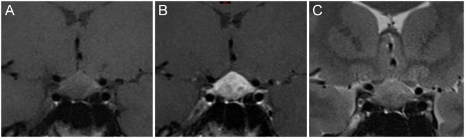
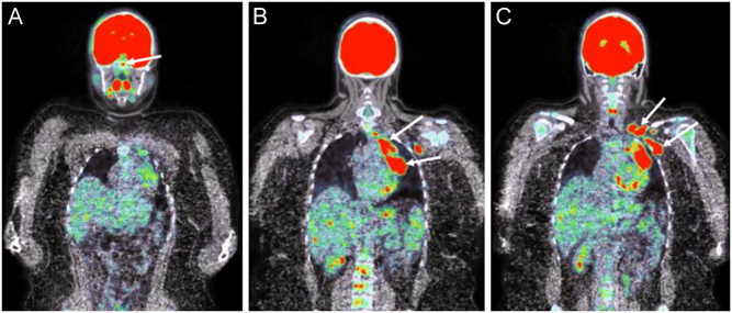
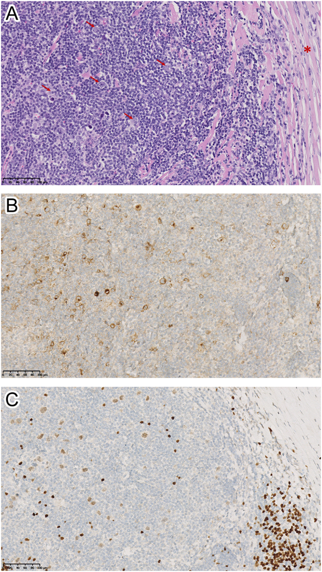
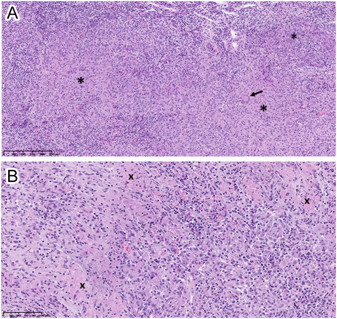
Summary: A 26-year-old woman presented with persistent headache and tiredness. Biological investigations disclosed a moderate inflammatory syndrome, low PTH-hypercalcemia and complete anterior hypopituitarism. A magnetic resonance imaging (MRI) of the pituitary gland was performed and revealed a symmetric enlargement with a heterogeneous signal. Ophthalmological examination showed an asymptomatic bilateral anterior and posterior uveitis, and a diagnosis of pituitary sarcoidosis was suspected. As the localization of lymphadenopathies on the fused whole-body FDG-PET/computerized tomography (CT) was not evoking a sarcoidosis in first instance, an excisional biopsy of a left supraclavicular adenopathy was performed showing classic nodular sclerosis Hodgkin's lymphoma (HL). A diagnostic transsphenoidal biopsy of the pituitary gland was proposed for accurate staging of the HL and surprisingly revealed typical granulomatous inflammation secondary to sarcoidosis, leading to the diagnosis of a sarcoidosis-lymphoma syndrome. The co-existence of these diseases constitutes a diagnostic challenge and we emphasize the necessity of exact staging of disease in order to prescribe adequate treatment.

Learning points: The possibility of a sarcoidosis-lymphoma syndrome, although rare, should be kept in mind during evaluation for lymphadenopathies. In the case of such association, lymphoma usually occurs after sarcoidosis. However, sarcoidosis and lymphoma can be detected simultaneously and development of sarcoidosis in a patient with previous lymphoma has also been reported. An accurate diagnosis of the disease and the respective organ involvements, including biopsy, is necessary in order to prescribe adequate treatment.

Keywords: 2019; 25-hydroxyvitamin-D3; ACTH; Adolescent/young adult; Belgium; C-reactive protein; CT scan; Chemotherapy; Cortisol; Cortisol (9am); Dacarbazine; FSH; FT4; Fatigue; Female; General practice; Glucocorticoids; Haematoxylin and eosin staining; Headache; Histopathology; Hydrocortisone; Hypercalcaemia; Hypopituitarism; IGF1; Immunohistochemistry; LH; Levothyroxine; Lymphadenitis; Lymphadenopathy; MRI; Methylprednisolone; New disease or syndrome: presentations/diagnosis/management; PET scan; PTH; Pituitary; Sarcoidosis; September; TSH; Thyroxine (T4); White; X-ray.


Figures

Figure 1
Figure 1
(A, B and C). Coronal T1-weighted view of the brain MRI (panel A) showing a symmetric enlargement of the pituitary gland with a marked and heterogeneous signal enhancement after gadolinium injection (panel B). Coronal T2-weighted view of the pituitary lesion is shown on panel C.
Figure 2
Figure 2
(A, B and C). Fused FDG-PET/CT scan showing enhanced metabolic signal at the level of the pituitary gland (arrow, panel A), as well as large metabolic unilateral hilar and mediastinal lymphadenopathies (panel B), as well as left supraclavicular and axillar adenopathies (arrows, panel C)
Figure 3
Figure 3
(A, B and C). Pathological examination of the supraclavicular lymph node biopsy specimen. Panel A (magnification ×20): hematoxylin-eosin staining showing a lymph node structure distorted by fibrotic bands (red asterisk) surrounding nodular reactive cell infiltrate composed of variable proportions of lymphocytes, histiocytes, eosinophils, and plasma cells and containing scattered large, malignant lymphoid cells. The abnormal large cells include bi-nucleated Reed-Sternberg cells (indicated by the red arrows), mononucleated Hodgkin cells and mummified cells. Panels B and C (magnification ×20): immunohistochemistry staining showing that the malignant lymphoid cells expressed CD30, a marker for HL (panel B), and PAX5, a transcription factor specific of B-cell lineage (panel C).
Figure 4
Figure 4
(A and B). Pathological examination of the pituitary biopsy specimen. Panel A (magnification ×10): hematoxylin and eosin staining showing diffuse lymphocytic inflammation with granuloma formation (indicated by the black asterisks). A multinucleated giant cell is also seen (arrow). Panel B (magnification x20): typical granulomatous inflammation with scattered areas of non-caseating necrosis (indicated by the black crosses). Normal pituitary tissue is not identified in the specimen and there are no histological signs of lymphoma.

References

    1. Cohen PR, Kurzrock R. Sarcoidosis and malignancy. Clinics in Dermatology 2007. 326–333. (10.1016/j.clindermatol.2007.03.010) - DOI - PubMed
    1. Ji J, Shu X, Li X, Sundquist K, Sundquist J, Hemminki K. Cancer risk in hospitalized sarcoidosis patients: a follow-up study in Sweden. Annals of Oncology 2009. 1121–1126. (10.1093/annonc/mdn767) - DOI - PubMed
    1. Anthony J, Esper GJ, Ioachimescu A. Hypothalamic pituitary sarcoidosis with vision loss and hypopituitarism: case series and literature review. Pituitary 2016. 19–29. (10.1007/s11102-015-0678-x) - DOI - PubMed
    1. Himiz K, Foyle A, Wilke D, Burrell S, Brownstone R, Ago C, Pahil R, Couban S. Intracranial presentation of systemic Hodgkin’s disease. Leukemia and Lymphoma 2004. 1667–1671. (10.1080/10428190410001673409) - DOI - PubMed
    1. Guermazi A, Brice P, de Kerviler E, Fermé C, Hennequin C, Meignin V, Frija J. Extranodal Hodgkin disease: spectrum of disease. RadioGraphics 2001. 161–179. (10.1148/radiographics.21.1.g01ja02161) - DOI - PubMed