

Doxorubicin-induced skeletal muscle atrophy: Elucidating the underlying molecular pathways
- PMID: 31600860
- PMCID: PMC7317437
- DOI: 10.1111/apha.13400
Doxorubicin-induced skeletal muscle atrophy: Elucidating the underlying molecular pathways
Abstract
Aim: Loss of skeletal muscle mass is a common clinical finding in cancer patients. The purpose of this meta-analysis and systematic review was to quantify the effect of doxorubicin on skeletal muscle and report on the proposed molecular pathways possibly leading to doxorubicin-induced muscle atrophy in both human and animal models.
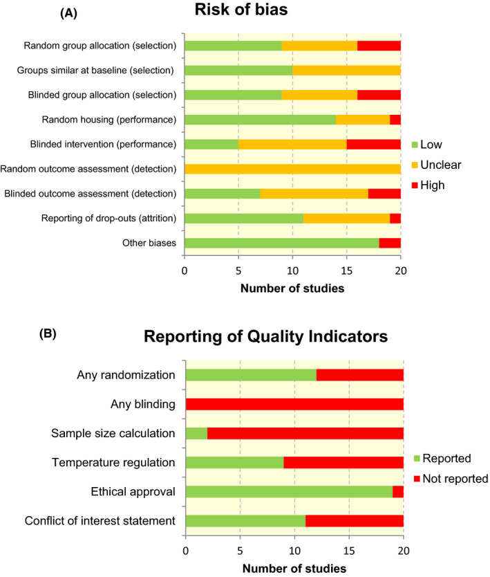
Methods: A systematic search of the literature was conducted in PubMed, EMBASE, Web of Science and CENTRAL databases. The internal validity of included studies was assessed using SYRCLE's risk of bias tool.
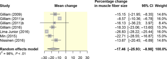
Results: Twenty eligible articles were identified. No human studies were identified as being eligible for inclusion. Doxorubicin significantly reduced skeletal muscle weight (ie EDL, TA, gastrocnemius and soleus) by 14% (95% CI: 9.9; 19.3) and muscle fibre cross-sectional area by 17% (95% CI: 9.0; 26.0) when compared to vehicle controls. Parallel to negative changes in muscle mass, muscle strength was even more decreased in response to doxorubicin administration. This review suggests that mitochondrial dysfunction plays a central role in doxorubicin-induced skeletal muscle atrophy. The increased production of ROS plays a key role within this process. Furthermore, doxorubicin activated all major proteolytic systems (ie calpains, the ubiquitin-proteasome pathway and autophagy) in the skeletal muscle. Although each of these proteolytic pathways contributes to doxorubicin-induced muscle atrophy, the activation of the ubiquitin-proteasome pathway is hypothesized to play a key role. Finally, a limited number of studies found that doxorubicin decreases protein synthesis by a disruption in the insulin signalling pathway.
Conclusion: The results of the meta-analysis show that doxorubicin induces skeletal muscle atrophy in preclinical models. This effect may be explained by various interacting molecular pathways. Results from preclinical studies provide a robust setting to investigate a possible dose-response, separate the effects of doxorubicin from tumour-induced atrophy and to examine underlying molecular pathways. More research is needed to confirm the proposed signalling pathways in humans, paving the way for potential therapeutic approaches.
Keywords: doxorubicin; mitochondrial dysfunction; muscle atrophy; reactive oxygen species; skeletal muscle; ubiquitin-proteasome pathway.
© 2019 The Authors. Acta Physiologica published by John Wiley & Sons Ltd on behalf of Scandinavian Physiological Society.
Conflict of interest statement
All authors declare that they have no conflict of interest.
Figures


References
-
- Carvalho C, Santos RX, Cardoso S, et al. Doxorubicin: the Good, the bad and the ugly effect. Curr Med Chem. 2009;16:3267‐3285. - PubMed
-
- Tacar O, Sriamornsak P, Dass CR. Doxorubicin: an update on anticancer molecular action, toxicity and novel drug delivery systems. J Pharm Pharmacol. 2013;65:157‐170. - PubMed
-
- Baracos VE, Martin L, Korc M, Guttridge DC, Fearon KCH. Cancer‐associated cachexia. Nat Rev Dis Prim. 2018;4:1‐18. - PubMed
Publication types
MeSH terms
Substances
LinkOut - more resources
Full Text Sources
Other Literature Sources
