. 2019 Dec;371(3):642-651.
doi: 10.1124/jpet.119.261792. Epub 2019 Oct 10.

Therapeutic Effects of Nrf2 Activation by Bardoxolone Methyl in Chronic Heart Failure

Affiliations

Therapeutic Effects of Nrf2 Activation by Bardoxolone Methyl in Chronic Heart Failure

Changhai Tian et al. J Pharmacol Exp Ther. 2019 Dec.

Abstract

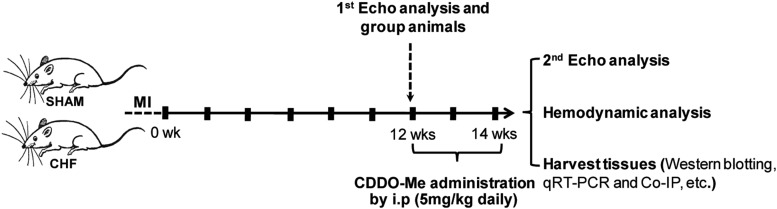
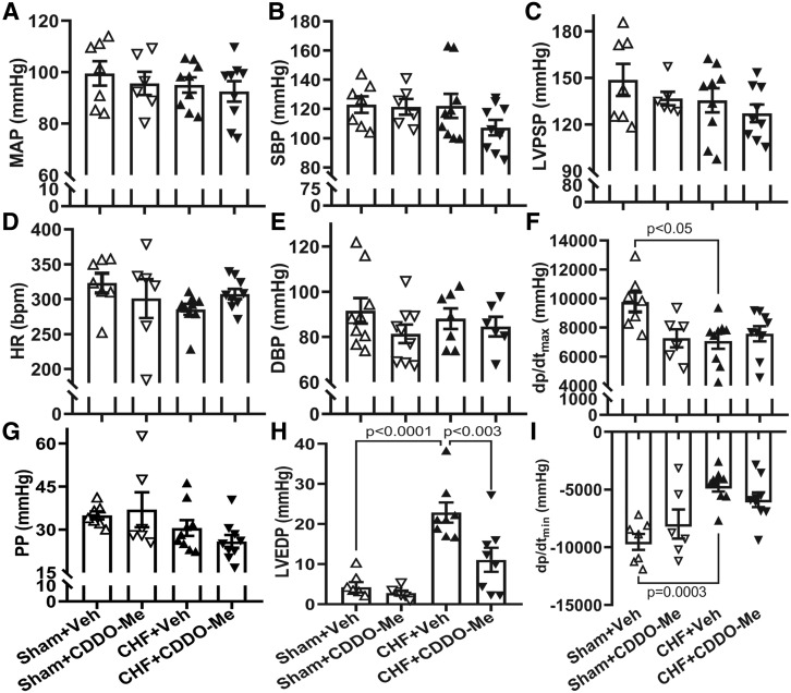
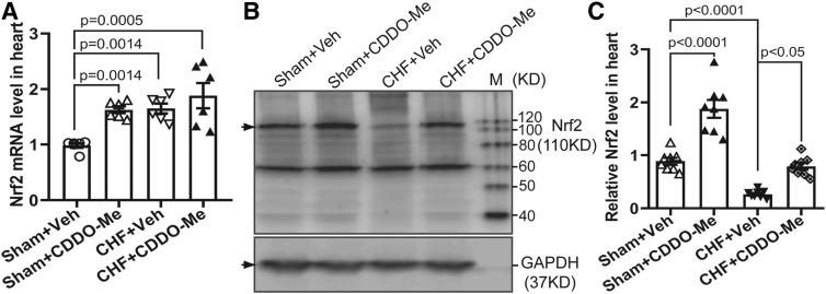
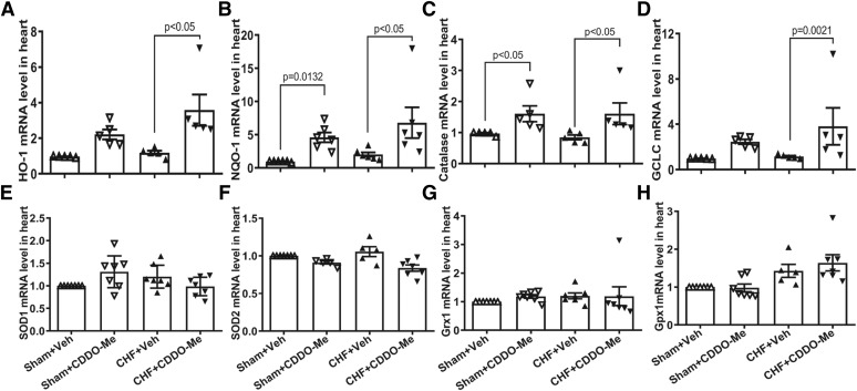
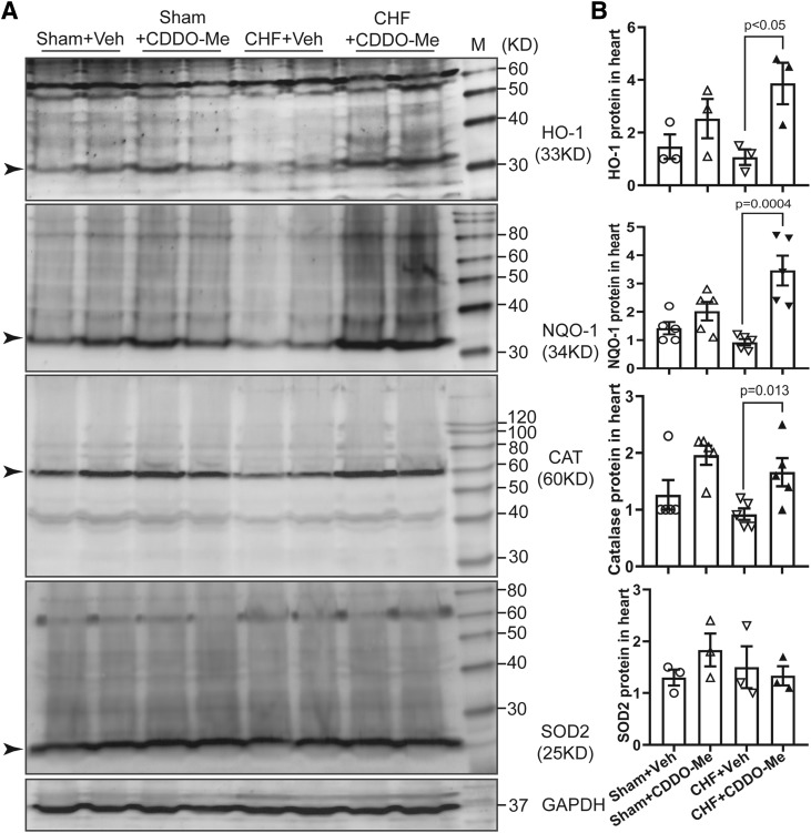
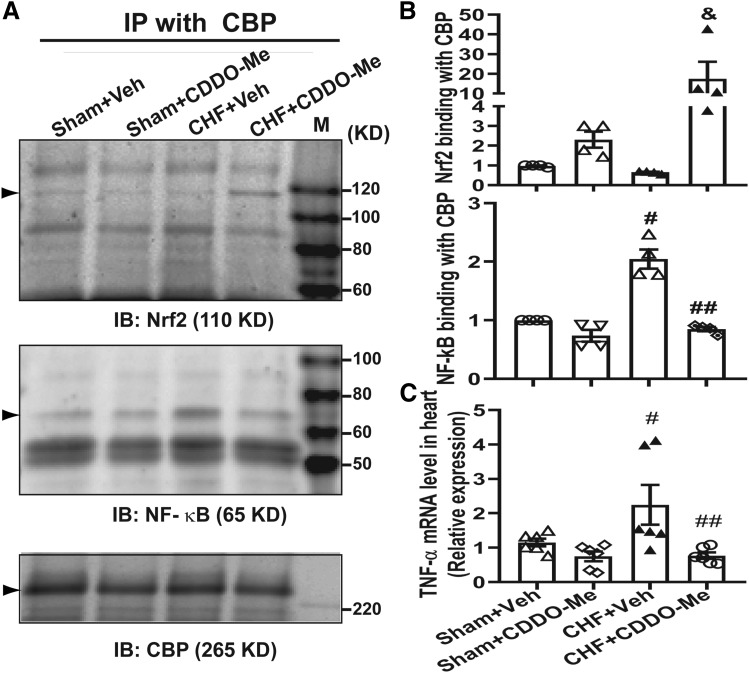
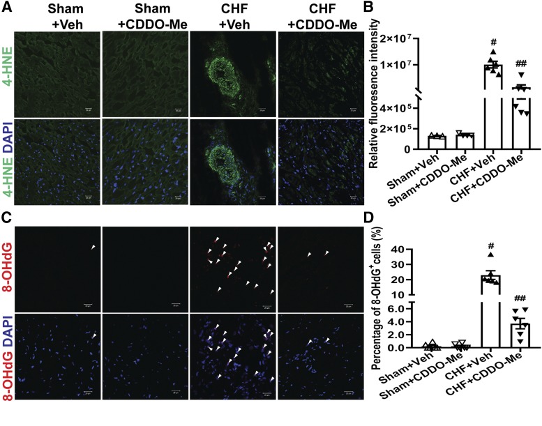
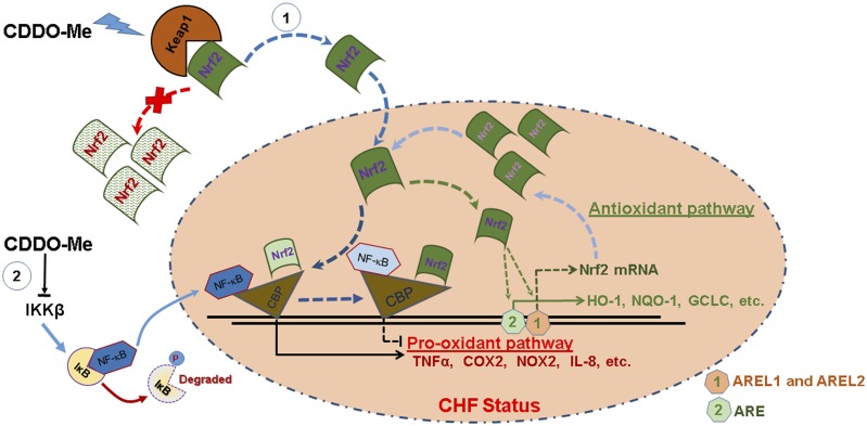
Oxidative stress plays an important role in the pathogenesis of chronic heart failure (CHF) in many tissues. Increasing evidence suggests that systemic activation of nuclear factor (erythroid-derived 2)-like 2 (Nrf2) signaling can protect against postinfarct cardiac remodeling by reducing oxidative stress. However, it remains to be elucidated if Nrf2 activation exerts therapeutic effects in the CHF state. Here, we investigated the beneficial hemodynamic effects of bardoxolone methyl (2-Cyano-3,12-dioxooleana-1,9(11)-dien-28-oic acid methyl ester, CDDO-Me), a pharmacological activator of Nrf2, in a rodent model of CHF. Based on echocardiographic analysis, rats at 12 weeks post-myocardial infarction (MI) were randomly split into four groups. CDDO-Me (5 mg/kg, i.p.) was administered daily for another 2 weeks in sham and CHF rats and compared with vehicle treatment. Echocardiographic and hemodynamic analysis suggest that short-term CDDO-Me administration increased stroke volume and cardiac output in CHF rats and decreased left ventricle end-diastolic pressure. Molecular studies revealed that CDDO-Me-induced cardiac functional improvement was attributed to an increase of both Nrf2 transcription and translation, and a decrease of oxidative stress in the noninfarcted areas of the heart. Furthermore, CDDO-Me reduced NF-κB binding and increased Nrf2 binding to the CREB-binding protein, which may contribute to the selective increase of Nrf2 downstream targets, including NADPH Oxidase Quinone 1, Heme Oxygenase 1, Catalase, and Glutamate-Cysteine Ligase Catalytic Subunit, and the attenuation of myocardial inflammation in CHF rats. Our findings suggest that Nrf2 activation may provide beneficial cardiac effects in MI-mediated CHF. SIGNIFICANCE STATEMENT: Chronic heart failure (CHF) is the leading cause of death among the aged worldwide. The imbalance between pro- and antioxidant pathways is a determinant in the pathogenesis of CHF. Systemic activation of Nrf2 and antioxidant protein signaling by bardoxolone methyl may have beneficial effects on cardiac function and result in improvements by enhancing antioxidant enzyme expression and attenuating myocardial inflammation.


Figures

Fig. 1.
Fig. 1.
Outline of the experimental protocol. Rats were allowed to recover for 12 weeks after thoracic surgery. At this time, an echocardiogram was taken, and treatment with either vehicle or CDDO-Me was begun for 2 weeks. At 14 weeks post thoracic surgery, a second echocardiogram was taken, and hemodynamic measurements were determined and tissues taken for biochemical analyses. Co-IP, coimmunoprecipitation; qRT-PCR, quantitative reverse-transcription polymerase chain reaction; i.p, intraperitoneal; Echo, Echocardiogram.
Fig. 2.
Fig. 2.
Hemodynamic data from CHF rats treated with CDDO-Me. Blood pressure (A); systolic pressure (SBP) (B); left ventricular peak-systolic pressure (LVPSP) (C); heart rate (HR) (D); diastolic blood pressure (DBP) (E), and pulse pressure (PP) (G). No significant difference was observed between different groups. Left-ventricular maximum rate of pressure increase (dp/dtmax) and decrease (dp/dtmin) were significantly decreased in CHF rats treated with vehicle compared with that in sham rats treated with vehicle (F and I), and LVEDP was significantly increased in Veh-treated CHF rats and partially restored after CDDO-Me treatment (H). Sham-Veh (n = 7), sham–CDDO-Me (n = 6), CHF-Veh (n = 8), CHF-CDDO-Me (n = 8) (±S.E.M.). MAP, mean arterial pressure.
Fig. 3.
Fig. 3.
CDDO-Me elevated both mRNA and protein levels of Nrf2 in the infarcted heart. (A) Quantitative reverse-transcription polymerase chain reaction data show CDDO-Me increases the Nrf2 transcription in the heart (n = 6, ±S.E.M.). Typical immunoblots (B) and pooled data (C) showing CDDO-Me induced the translational increase of Nrf2 in the noninfarcted left ventricle 2 weeks post CDDO-Me administration (n = 7, ±S.E.M.). GAPDH, glyceraldehyde-3-phosphate dehydrogenase.
Fig. 4.
Fig. 4.
Cardiac tissues from left ventricles were collected and then subjected to quantitative reverse-transcription polymerase chain reaction analysis with specific primers for HO-1 (A), NQO1 (B), catalase (C), glutamate-cysteine ligase (GCLC) (D), SOD1 (E), SOD2 (F), Grx1 (G), and Gpx1 (H). Glyceraldehyde-3-phosphate dehydrogenase (GAPDH) was used as an internal control (n = 5 to 6, ±S.E.M.).
Fig. 5.
Fig. 5.
Sham and CHF rats were treated with vehicle or CDDO-Me, respectively, for 2 weeks. The noninfarcted area of the left ventricles were collected and subjected to western blotting analysis for HO-1, NQO1, catalase, and SOD2. Glyceraldehyde-3-phosphate dehydrogenase (GAPDH) was used as a loading control (A); mean data are shown in (B) (±S.E.M.) as the ratio of each protein to GAPDH.
Fig. 6.
Fig. 6.
Left ventricular tissue was subjected to coimmunoprecipitation with CBP antibody and then western blotting analysis with Nrf2 and NF-kB (p65) antibody, respectively. Immunoblots (A) and pooled data (B) showing the enhanced binding of Nrf2 protein to CBP and reduced binding of NF-kB to CBP in the noninfarcted left ventricle 2 weeks post CDDO-Me administration (n = 4, ±S.E.M.). &P < 0.05 vs. CHF+Veh; #P < 0.0001 vs. sham+Veh; ##P < 0.0001 vs. CHF+Veh. (C) The noninfarcted region of the left ventricle was collected and then subjected to RNA extraction and quantitative reverse-transcription polymerase chain reaction analysis with specific primers for TNF-α. Quantitative reverse-transcription polymerase chain reaction data show CDDO-Me inhibits the transcription of TNF-α in the infarcted heart compared with that in the Veh-treated CHF group (n = 6, ±S.E.M.). #P < 0.05 vs. sham+Veh; ##P < 0.05 vs. CHF+Veh. IB, XXX; IP, immunoprecipitation; IB, immunoblotting.
Fig. 7.
Fig. 7.
CDDO-Me decreases oxidative stress of the heart. Oxidative stress in the left ventricles of hearts was determined in sham and CHF rats (12 weeks post MI) that were treated with vehicle or CDDO-Me for an additional 2 weeks, respectively. (A) Representative confocal microscopic images of the left ventricle with 4-HNE staining. 4-HNE–positive is shown in green. (B) The relative fluorescence intensities were quantified by ImageJ software (NIH) (sham+Veh: n = 3; sham+CDDO-Me: n = 4; CHF+Veh and CHF+CDDO-Me: n = 6). #P < 0.0001 vs. sham+Veh; ##P < 0.0001 vs. CHF+Veh. (C) Representative confocal microscopic images of the left ventricle 8-OHdG staining. 8-OHdG–positive is shown in red. (D) The percentage of 8-OHdG+ cells was quantified (sham+Veh and sham+CDDO-Me: n = 4; CHF+Veh and CHF+CDDO-Me: n = 6). #P < 0.0001 vs. sham+Veh; ##P < 0.0001 vs. CHF+Veh. Nuclei are shown in blue (DAPI, 4′,6-diamidino-2-phenylindole). Original magnification, 400×. All images were processed with the same confocal settings.
Fig. 8.
Fig. 8.
A schematic diagram of the potential mechanisms for CDDO-Me–mediated Nrf2 activation in CHF. On the one hand, CDDO-Me promotes the dissociation of Nrf2 from Kelch-like ECH-associated protein 1 (Keap1; a natural Nrf2 inhibitor) in the cytosol, thus translocating to the nucleus where enhanced binding of Nrf2 to CBP promotes pro-antioxidant pathways, and reduced binding of NF-κB to CBP results in the inhibition of the pro-oxidant pathway in CHF. At the same time, Nrf2 binds to its own promoter at AREL1 and AREL2 sites to amplify its antioxidative effects by increasing its own transcription. On the other hand, CDDO-Me may inhibit the binding of NF-κB to DNA and subsequent transcriptional activation through the direct interaction with IκB kinase (IKK) β to prevent NF-κB p65 nuclear translocation. AREL1 and AREL2, Antioxidant Response Element (ARE)-Like Sequence 1 and 2.

References

    1. Appendino G, Belcaro G, Cornelli U, Luzzi R, Togni S, Dugall M, Cesarone MR, Feragalli B, Ippolito E, Errichi BM, et al. (2011) Potential role of curcumin phytosome (Meriva) in controlling the evolution of diabetic microangiopathy. A pilot study. Panminerva Med 53 (3 Suppl 1):43–49. - PubMed
    1. Ashrafian H, Czibik G, Bellahcene M, Aksentijević D, Smith AC, Mitchell SJ, Dodd MS, Kirwan J, Byrne JJ, Ludwig C, et al. (2012) Fumarate is cardioprotective via activation of the Nrf2 antioxidant pathway. Cell Metab 15:361–371. - PMC - PubMed
    1. Bai Y, Chen Q, Sun YP, Wang X, Lv L, Zhang LP, Liu JS, Zhao S, Wang XL. (2017) Sulforaphane protection against the development of doxorubicin-induced chronic heart failure is associated with Nrf2 Upregulation. Cardiovasc Ther 35:e12277. - PubMed
    1. Bai Y, Wang X, Zhao S, Ma C, Cui J, Zheng Y. (2015) Sulforaphane protects against cardiovascular disease via Nrf2 activation. Oxid Med Cell Longev 2015:407580. - PMC - PubMed
    1. Bhimaraj A, Tang WH. (2012) Role of oxidative stress in disease progression in Stage B, a pre-cursor of heart failure. Heart Fail Clin 8:101–111. - PMC - PubMed

Publication types