. 2019 Oct 11;366(6462):eaav5728.
doi: 10.1126/science.aav5728.

Migratory DCs activate TGF-β to precondition naïve CD8+ T cells for tissue-resident memory fate

Affiliations

Migratory DCs activate TGF-β to precondition naïve CD8+ T cells for tissue-resident memory fate

Vinidhra Mani et al. Science. .

Abstract

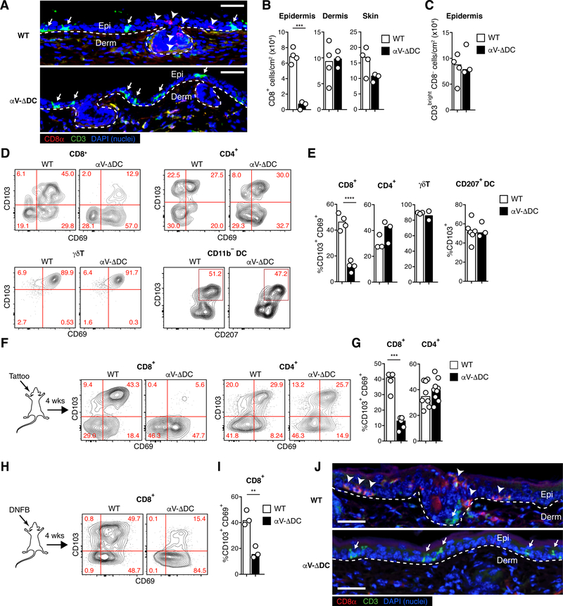
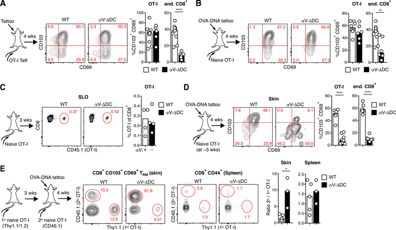
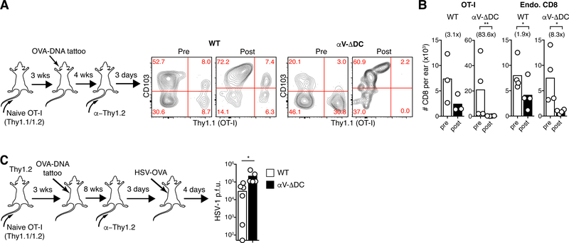
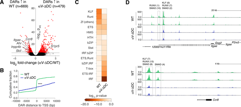
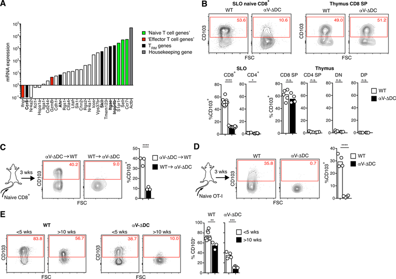
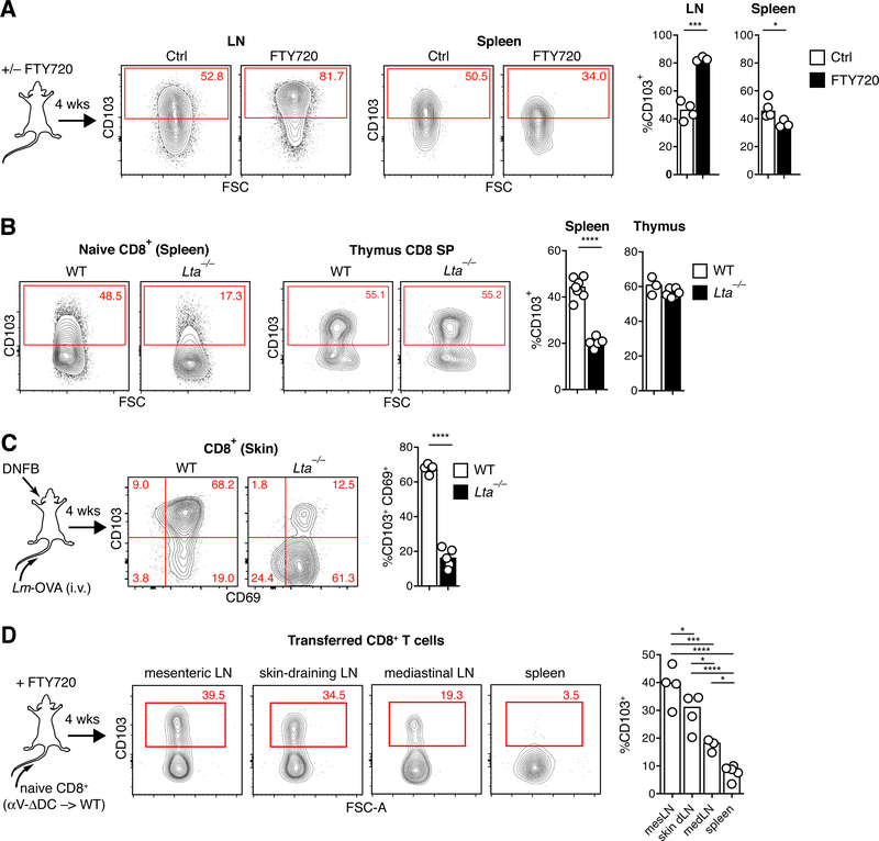
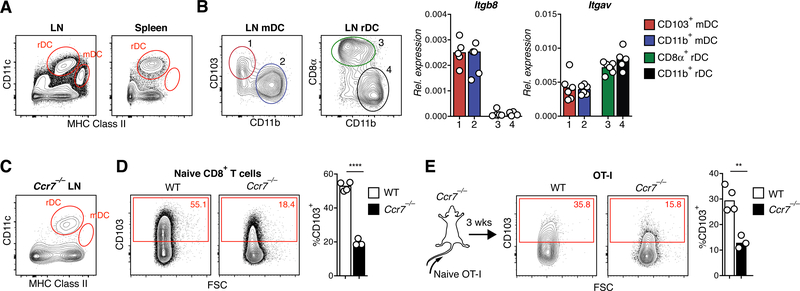
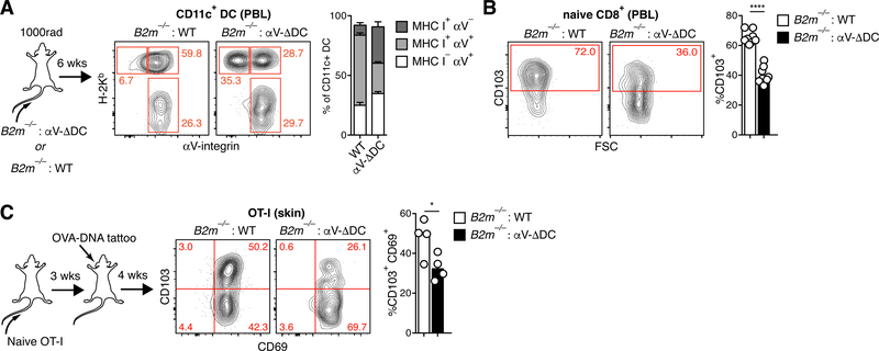
Epithelial resident memory T (eTRM) cells serve as sentinels in barrier tissues to guard against previously encountered pathogens. How eTRM cells are generated has important implications for efforts to elicit their formation through vaccination or prevent it in autoimmune disease. Here, we show that during immune homeostasis, the cytokine transforming growth factor β (TGF-β) epigenetically conditions resting naïve CD8+ T cells and prepares them for the formation of eTRM cells in a mouse model of skin vaccination. Naïve T cell conditioning occurs in lymph nodes (LNs), but not in the spleen, through major histocompatibility complex class I-dependent interactions with peripheral tissue-derived migratory dendritic cells (DCs) and depends on DC expression of TGF-β-activating αV integrins. Thus, the preimmune T cell repertoire is actively conditioned for a specialized memory differentiation fate through signals restricted to LNs.


Conflict of interest statement

Competing Interests:

The authors declare no competing interests

Figures

Fig. 1.
Fig. 1.. Defective epidermal TRM cell formation in absence of αV integrins on DCs.
(A) Histological cross-sections from ear skin of 8–10 week-old αV-ΔDC and WT littermate control mice. Arrowheads indicate CD8+ CD3dim TRM cells in the epidermis of WT, but not αV-ΔDC mice, whereas arrows indicate CD3bright DETCs present in both strains. Dashed lines indicate the dermal–epidermal border. Scale bar = 50 μm. Epi: epithelium, Derm: dermis. (B, C) Density of CD8+ T cells in epidermis, dermis, and total skin (B) and of CD3bright CD8 T cells in epidermis (C). Data are means and replicates and representative of two independent experiments. Each symbol represents the mean of 3–4 values per animal obtained from different histological sections. (D, E) Expression of the tissue residence marker CD103 (as well as CD69 on T cells) on indicated immune cell subsets (Thy1+ CD8β+ T cells, Thy1+ CD4+ T cells, Thy1+ TCRd+ γΔT cells, CD11c+ MHC II+ CD11b DCs) from skin of αV-ΔDC and WT littermate control mice. Data are means and replicates and representative of two independent experiments. (F-I) Expression of CD69 and CD103 on skin CD8+ or CD4+ T cells 4 weeks after sterile inflammation induced by mechanical irritation with a tattooing device (F, G) or topical DNFB treatment (H, I) in 8–10-week-old mice. Data are means and replicates and representative of five (F, G) or three (H, I) independent experiments. (J) Ear skin 4 weeks after DNFB treatment. Note the enrichment of CD8β+ CD3dim eTRM cells (arrow-heads) in WT mice, but their absence in the epithelium of αV-ΔDC, which is instead still populated by CD3bright DETC (arrows). Data are representative of two animals per group. Scale bar = 50 μm. **/***/****: p<0.01/p<0.001/p<0.0001 (Two-tailed unpaired Student’s t-tests in (B), (C), (E), (G), (I)).
Fig. 2.
Fig. 2.. DC-expressed αV-integrins condition naive CD8+ T cells for epithelial TRM cell formation prior to vaccine-induced activation.
(A) 106 Thy1.1/2 congenic ex vivo activated OT-I effector cells were intravenously injected into Thy1.2 αV-ΔDC or WT recipients, whose ears were simultaneously inflamed through tattoo injury. After 4 weeks, CD69 and CD103 expression was assessed on transferred OT-I and host CD8+ T cells in skin. Data are means and replicates and representative of six independent experiments. (B) 105 CD44lo CD62Lhi naive OT-I cells were adoptively transferred into αV-ΔDC or WT recipients, whose ears were simultaneously tattooed with OVA-encoding plasmid DNA to prime OT-I in skin-draining LNs. After 4 weeks, CD69 and CD103 expression was assessed on transferred OT-I and host CD8+ T cells in skin. Data are means and replicates and representative of five independent experiments. (C, D) 106 CD44lo CD62Lhi naive OT-I cells were adoptively transferred into αV-ΔDC or WT recipients. Three weeks later, OT-I cell frequency in pooled LNs and spleen (SLO) was determined in some animals (C), while the remaining animals were vaccinated by ear tattoo with OVA-encoding plasmid DNA (D). After 4 weeks, CD69 and CD103 expression was assessed on transferred OT-I and host CD8+ T cells in skin. Data are means and replicates and representative of four independent experiments. (E) 106 Thy1.1/2 congenic CD44lo CD62Lhi naive OT-I cells (1° OT-I) were adoptively transferred into αV-ΔDC or WT recipients. Three weeks later, a second batch of 105 CD45.1 congenic OT-I cells (2° OT-I) was transferred into the same recipients and mice were vaccinated by ear tattoo with OVA-encoding plasmid DNA. After 4 weeks, the ratios of OT-I derived from the second and the first injected batch was assessed in the pools of CD69+ CD103+ TRM cells in skin and of CD44+ CD8+ T cells in spleen. Data are means and replicates and representative of two independent experiments. */**/****: p<0.05/p<0.01/p<0.0001 (Two-tailed unpaired Student’s t-tests in (A-E)).
Fig. 3.
Fig. 3.. Residual eTRM cells formed in absence of DC-expressed αV-integrins are unstable and provide reduced antiviral protection.
(A-B) 106 CD44lo CD62Lhi naive OT-I cells were adoptively transferred into αV-ΔDC or WT recipients. Three weeks later, animals were vaccinated by ear tattoo with OVA-encoding plasmid DNA. After an additional 4 weeks, circulating T cells were depleted with α-Thy1.2 mAbs, and then, 3 d later, the expression of CD103 (A) and absolute numbers in skin of total CD8+ T cells (B) derived from OT-I (left) or from polyclonal endogenous cells (right) were determined. Number in parentheses indicate fold decrease in αV-ΔDC mice Data are means and replicates and representative of two independent experiments. (C) Mice were seeded with OT-I eTRM cells as in (A), but in the flank. Eight weeks later, circulating T cells were depleted, and an additional 3 d later, animals were exposed at vaccinated skin sites with HSV-OVA. Four days later, local viral titers were determined in skin. Data are medians and biological replicates, each replicate representing the mean of a technical triplicate, and representative of two independent experiments. */**: p<0.05/p<0.01 (Two-tailed paired Student’s t-tests in (B), *: p<0.05 (Mann–Whitney U test in (C)).
Fig. 4.
Fig. 4.. Resting naive CD8+ T cells are epigenetically conditioned by TGF-β signals in secondary lymphoid tissues.
(A) Volcano plot of chromatin accessibility changes between CD44lo naive CD8+ T cells from WT and αV-ΔDC mice, considering merged DARs. (B) Cumulative distributions of distances from DARs in cells from WT (green) and αV-ΔDC (blue) animals to the closest transcription start site. The two-sample Kolmogorov–Smirnov was used to compare cumulative distributions. (C) Enrichment of indicated transcription factor binding motif families in DARs. (D) Normalized chromatin accessibility near the Itgae (top) and Ccr8 (bottom) loci. The rectangles mark detected DARs. All analyses were performed on 2 mice per group with similar results. ****: p<0.0001 (Two-sample Kolmogorov–Smirnov test in (B)).
Fig. 5.
Fig. 5.. CD103 is actively maintained on naive CD8+ T cells through αV-integrin-expressing DCs.
(A) ImmGen database RNA-seq gene expression analysis of naive CD8+ T cells. Green bars denote selected genes known to be expressed at the protein level in naive T cells, red bars denote selected genes known to be expressed at very low levels or not to be expressed in naive T cells. Black bars denote skin eTRM cell genes having greater accessibility in naive CD8+ T cells from WT as compared to αV-ΔDC mice. White bars denote other skin eTRM cell genes not differentially accessible in cells from WT compared to αV-ΔDC mice. (B) Expression of CD103 on CD44lo CD62Lhi naive CD4+ and CD8+ T cells (pooled from LNs and spleen) and on thymocyte subsets from αV-ΔDC and WT littermate control mice. Data are means and replicates and representative of four independent experiments. (C) 106 naive polyclonal CD8+ T cells from αV-ΔDC were adoptively transferred into CD45.1 congenic C57BL/6 mice or vice versa and re-isolated 3 weeks later from pooled LNs and spleens for analysis of CD103 expression. Data are means and replicates and representative of three independent experiments. (D) 106 naive OT-I T cells were adoptively transferred into αV-ΔDC or WT hosts and isolated from pooled LNs and spleens 3 weeks later for analysis of CD103 expression. Data are means and replicates and representative of three independent experiments. (E) CD103 expression on CD44lo naive CD8+ T cells from the peripheral blood of WT and αV-ΔDC mice at less than 5 or more than 10 wks of age. Data are means and replicates. */**/***/****: p<0.05/p<0.01/p<0.001/p<0.0001 (Two-tailed unpaired Student’s t-tests in (B-E)).
Fig. 6.
Fig. 6.. Naive CD8+ T cell preconditioning occurs in lymph nodes, but not spleens.
(A) C57BL/6 mice were treated with FTY720 to block lymphocyte egress from all lymphoid tissues. After 4 weeks of continued treatment, naive CD8+ T cells in LNs and spleen were analyzed for CD103 expression. Data are means and replicates and representative of six independent experiments. (B) CD103 expression in naive CD8+ T cells in spleens and thymi of WT and Lta−/− mice. Data are means and replicates and representative of two independent experiments. (C) Frequency of CD69+ CD103+ eTRM cells in skin 4 weeks after ‘prime and pull’ immune challenge through i.v. injection with OVA-expressing Listeria monocytogenes to produce a circulating TEFF cell pool (“prime”) followed by DNFB+ treatment of ear skin (“pull”) of WT an Lta−/− mice. Data are means and replicates and representative of four independent experiments. (D) CD103 expression of naive CD8+ T cells from αV-ΔDC donors in indicated SLOs of continuously FTY720-treated WT hosts 4 weeks following adoptive transfer. Data are means and replicates and representative of two independent experiments. */**/***/****: p<0.05/p<0.01/p<0.001/p<0.0001 (Two-tailed unpaired Student’s t-tests in (A-C), One-way ANOVA in (D)).
Fig. 7.
Fig. 7.. αVβ8-expressing migratory DCs precondition naive CD8+ T cells for epithelial TRM cell formation in skin
(A) CD11cint MHC IIhi migratory DCs in LNs are absent in spleens of C57BL/6 mice. (B) CD103+ and CD11b+ subsets of migratory DCs as well as CD8+ and CD11b+ subsets of resident DCs were purified by FACS and analyzed for expression of αV and β8 integrin mRNA by RT-qPCR. Data are means and replicates and representative of two independent experiments. (C) CD11cint MHC IIhi migratory DCs are absent in LNs of Ccr7−/− mice. (D) CD103 expression in naive CD8+ T cells in pooled LNs and spleen of Ccr7−/− mice. Data are means and replicates and representative of four independent experiments. (E) 106 naive OT-I T cells were adoptively transferred into Ccr7−/− or C57BL/6 hosts. CD103 expression on naive OT-I cells from pooled LNs and spleen was analyzed 3 weeks later. Data are means and replicates and representative of two independent experiments. **/****: p<0.01/p<0.0001 (Two-tailed unpaired Student’s t-tests in (D-E)).
Fig. 8.
Fig. 8.. eTRM cell preconditioning of naive CD8+ T cells occurs through homeostatic MHC I-dependent DC interactions.
(A) Lethally irradiated CD45.1 congenic mice were injected with 1:1 mixtures of either WT or αV-ΔDC and MHC I-deficient B2m−/− bone marrow cells. Eight weeks later, the expression of H-2Kb and αV on CD45.1+ CD11+ cells in peripheral blood was assessed to determine the frequency of cells that express either one of both proteins. Data are means ± SEM (n=8 for WT and n=6 for αV-ΔDC) and representative of four independent experiments. (B) CD103 expression on naive CD8+ T cells in the peripheral blood of αV-ΔDC : B2m−/− mixed BMCs, in which expression of αV and MHC I on DCs is segregated. Data are means and replicates and representative of two independent experiments. (C) 106 naive OT-I cells were adoptively transferred into BMCs. Three weeks later, mice were vaccinated by ear tattoo with OVA-encoding plasmid DNA. After an additional 4 weeks, CD69 and CD103 expression was assessed on transferred OT-I T cells in skin. Data are means and replicates and representative of two independent experiments. */****: p<0.05/p<0.0001 (Two-tailed unpaired Student’s t-tests in (B-C)).

Comment in

References

    1. Masopust D, Soerens AG, Tissue-Resident T Cells and Other Resident Leukocytes. Annu. Rev. Immunol. 37, 521–546 (2019). - PMC - PubMed
    1. Mueller SN, Mackay LK, Tissue-resident memory T cells: Local specialists in immune defence. Nat. Rev. Immunol. 16, 79–89 (2016). - PubMed
    1. Gebhardt T, Palendira U, Tscharke DC, Bedoui S, Tissue-resident memory T cells in tissue homeostasis, persistent infection, and cancer surveillance. Immunol. Rev. 283, 54–76 (2018). - PubMed
    1. Gebhardt T et al., Memory T cells in nonlymphoid tissue that provide enhanced local immunity during infection with herpes simplex virus. Nat. Immunol. 10, 524–530 (2009). - PubMed
    1. Schumacher TN et al., Skin-resident memory CD8+ T cells trigger a state of tissue-wide pathogen alert. Science (80-. ). 346, 101–105 (2014). - PubMed

Publication types

Substances