Review
. 2019 Oct 11;366(6462):eaaw9997.
doi: 10.1126/science.aaw9997.

Axonal transport: Driving synaptic function

Affiliations
Review

Axonal transport: Driving synaptic function

Pedro Guedes-Dias et al. Science. .

Abstract

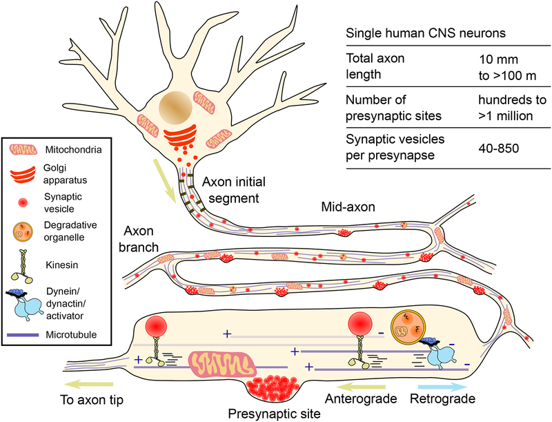
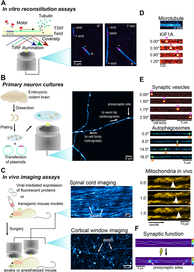
The intracellular transport system in neurons is specialized to an extraordinary degree, enabling the delivery of critical cargo to sites in axons or dendrites that are far removed from the cell center. Vesicles formed in the cell body are actively transported by kinesin motors along axonal microtubules to presynaptic sites that can be located more than a meter away. Both growth factors and degradative vesicles carrying aged organelles or aggregated proteins take the opposite route, driven by dynein motors. Distance is not the only challenge; precise delivery of cargos to sites of need must also be accomplished. For example, localized delivery of presynaptic components to hundreds of thousands of "en passant" synapses distributed along the length of a single axon in some neuronal subtypes provides a layer of complexity that must be successfully navigated to maintain synaptic transmission. We review recent advances in the field of axonal transport, with a focus on conceptual developments, and highlight our growing quantitative understanding of neuronal trafficking and its role in maintaining the synaptic function that underlies higher cognitive processes such as learning and memory.


Figures

Fig. 0.
Fig. 0.. Axonal transport drives cargo through extreme geometries.
Neurons in the human central nervous system (CNS) display highly complex axonal arbors that can branch thousands of times, reach hundreds of meters in total length, and contain hundreds of thousands of presynaptic sites distributed ‘en passant’. The axonal transport machinery is critical for synaptic function by delivering new synaptic vesicles to and removing aged organelles from presynaptic sites.
Fig. 1.
Fig. 1.. Axonal microtubule organization and polarity.
Stabilization of the microtubule “plus-end out” arrangement in the axon initial segment (AIS) is critical for axonal identity and likely acts as a template to establish the uniform polarity of the axonal microtubule network along the mid-axon.
Fig. 2.
Fig. 2.. Axonal microtubule diversity.
From numerous tubulin isoforms, to distinct nucleotide states and several PTMs, microtubule lattices can in theory display a large number of combinatorial arrangements. This diversity can presumably be shown either at the single microtubule level (multitude of tubulin isoforms, nucleotide states, and PTMs distributed across the same microtubule lattice), or at the microtubule population level (uniformity of tubulin isoforms, nucleotide state and PTM distribution across microtubule populations, possibly conferring these a specific function).
Fig. 3.
Fig. 3.. Axonal transport is regulated at multiple levels.
Despite having distinct biophysical properties, (A) kinesin and (D) dynein undergo similar transport phases. From an inactive unbound state, the motors bind to cargo and are recruited to the microtubule where they initiate active transport. MAPs, such as Tau or MAP7 can differentially affect motors and induce transport termination in a motor-specific manner. Kinesin-3 can sense the nucleotide state of the microtubule lattice and rapidly detaches upon encountering a dynamic GTP microtubule plus-end. Initiation (B) and termination (C) of synaptic vesicle precursor (SVP) transport along an axon of a live hippocampal neuron in culture expressing synaptophysin-mScarlet. SVPs are transported mostly in the anterograde direction by kinesin-3 and pause preferentially at presynaptic sites, which are enriched in dynamic GTP-rich microtubule plus-ends. Initiation (E) and termination (F) of autophagosome transport along the axon of a live cultured hippocampal neuron expressing LC3-GFP. Autophagosomes are mostly generated distally in the axon and driven by dynein in the retrograde direction towards the soma.
Fig. 4.
Fig. 4.. Range of imaging approaches to address questions in the field of axonal transport.
A wide range of imaging assays can be employed to investigate the dynamics of biological processes in systems with different levels of biological complexity. For example: A) Combining in vitro reconstitution assays with total internal reflection fluorescence (TIRF) microscopy allows the assessment of the dynamic behavior of single motors on microtubules. The ability to easily manipulate the system by adding or removing components enables the elucidation of how single factors (e.g. MAPs) affect motor activity or what are the minimum components required in the system to observe a specific dynamic behavior. Panels show the basic setup of an in vitro reconstitution assay and TIRF microscopy imaging of polymerization of fluorescently-labelled tubulin. B) Primary rodent CNS neurons in culture undergo a well-described polarized developmental process akin to what is observed in vivo. Neurons develop an axon and multiple dendrites, establish synaptic contacts with their neighbors, and fire action potentials spontaneously. The combined ability to easily manipulate the neurons genetically and pharmacologically, express fluorescent reporters and use imaging techniques with high spatial and temporal resolution such as spinning disk confocal microscopy, make this system ideal to investigate the mechanisms underlying organelle dynamics in a physiologically-relevant context. Panels show a basic protocol to image primary neuron cultures and a confocal microscopy image of hippocampal axons in culture expressing fluorescently-tagged synaptophysin. C) The development of Cre mouse lines, improved adeno-associated viral vectors, and the advent of CRISPR/Cas9 gene-editing techniques have enabled more straightforward strategies of genetically manipulating mice, generating transgenic mouse lines, and expressing fluorescent reporters even in a neuron sub-type-specific manner. Innovative surgical techniques, including spinal cord exposure and cortical window implementation allow long-term optical access to the spinal cord and superficial layers of the cortex. By employing two-photon microscopy techniques, it is thus possible to interrogate organelle dynamics and synaptic function in neurons in their native environment. Panels show a simple strategy to perform in vivo imaging assays, an image of axonal bundles in the spinal cord of a Thy1-YFP mouse, and a cholinergic axon in cortical layer I/II of a ChAT-Cre ROSA26-EGFPf mouse. D) TIRF imaging of a single dimeric KIF1A motor labeled with TMR actively moving on a fluorescently-labelled microtubule in an in vitro reconstitution assay. E) Synaptic vesicle precursors (labeled with synaptophyin-mScarlet) move mostly in the anterograde direction, while autophagosomes (labeled with LC3-GFP) show robust retrograde movement along the axons of live neurons in culture. Two-photon microscopy imaging of mitochondrial dynamics in spinal cord axons of a Thy1-MitoCFP mouse. F) VGLUT1-pHluorin signal in a primary hippocampal neuron before and after electrical-field stimulation. An increased repertoire of fluorescent pH and Ca2+ indicators are allowing the study of how a variety of processes and mechanisms regulating axonal transport affects synaptic fuhnction and neuronal activity.

References

    1. Chaaban S, Brouhard GJ, A microtubule bestiary: structural diversity in tubulin polymers. Molecular biology of the cell 28, 2924–2931 (2017). - PMC - PubMed
    1. Akhmanova A, Steinmetz MO, Control of microtubule organization and dynamics: two ends in the limelight. Nature reviews. Molecular cell biology 16, 711–726 (2015). - PubMed
    1. Nirschl JJ, Ghiretti AE, Holzbaur ELF, The impact of cytoskeletal organization on the local regulation of neuronal transport. Nature reviews. Neuroscience 18, 585–597 (2017). - PMC - PubMed
    1. Olenick MA, Holzbaur ELF, Dynein activators and adaptors at a glance. Journal of cell science 132, (2019). - PMC - PubMed
    1. Roy S, Seeing the unseen: the hidden world of slow axonal transport. The Neuroscientist : a review journal bringing neurobiology, neurology and psychiatry 20, 71–81 (2014). - PMC - PubMed

Publication types