. 2019 Oct 11;366(6462):203-210.
doi: 10.1126/science.aax3939.

Architecture of human Rag GTPase heterodimers and their complex with mTORC1

Affiliations

Architecture of human Rag GTPase heterodimers and their complex with mTORC1

Madhanagopal Anandapadamanaban et al. Science. .

Abstract

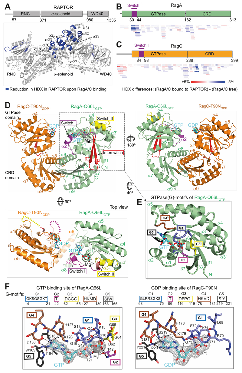
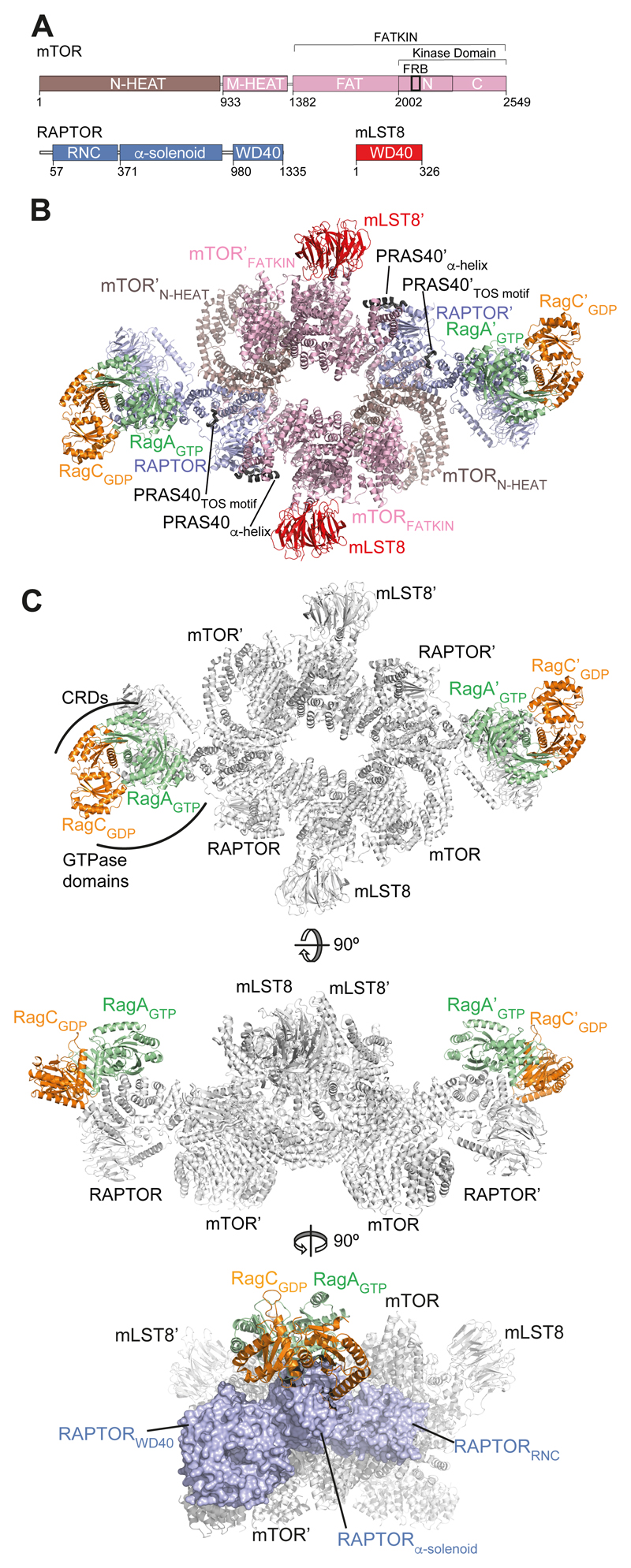
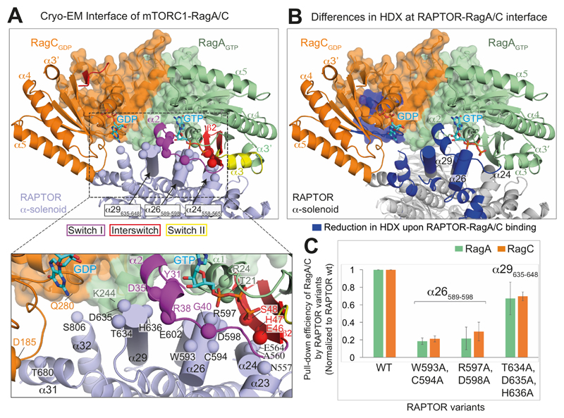
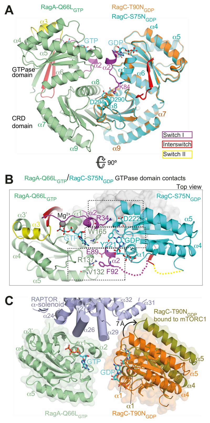
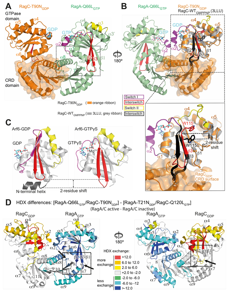
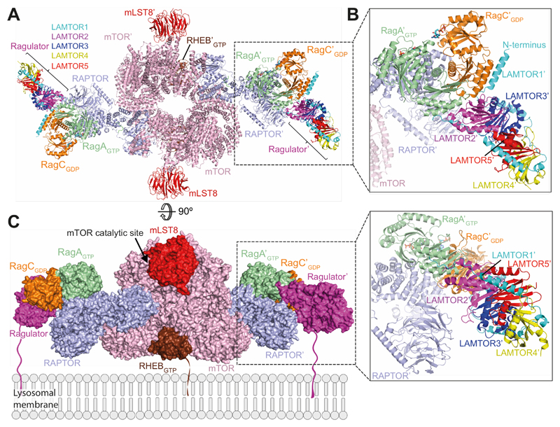
The Rag guanosine triphosphatases (GTPases) recruit the master kinase mTORC1 to lysosomes to regulate cell growth and proliferation in response to amino acid availability. The nucleotide state of Rag heterodimers is critical for their association with mTORC1. Our cryo-electron microscopy structure of RagA/RagC in complex with mTORC1 shows the details of RagA/RagC binding to the RAPTOR subunit of mTORC1 and explains why only the RagAGTP/RagCGDP nucleotide state binds mTORC1. Previous kinetic studies suggested that GTP binding to one Rag locks the heterodimer to prevent GTP binding to the other. Our crystal structures and dynamics of RagA/RagC show the mechanism for this locking and explain how oncogenic hotspot mutations disrupt this process. In contrast to allosteric activation by RHEB, Rag heterodimer binding does not change mTORC1 conformation and activates mTORC1 by targeting it to lysosomes.


Conflict of interest statement

Competing interests: Authors declare no competing interests.

Figures

Fig. 1
Fig. 1. The crystal structure of a RagA/C heterodimer and HDX-MS analysis of its interaction with RAPTOR.
(A) HDX-MS identified regions protected from HDX in the RAPTOR/RagA-Q66LGTP/RagCT90NGDP complex. Decreases in HDX (blue) of RAPTOR upon RagA/C binding are depicted on the RAPTOR structure (from PDB ID: 6BCX). (B, C) Differences in HDX for RagA-Q66LGTP (B) and RagC-T90NGDP (C) upon RAPTOR binding for all the peptides at 0.3 s in D2O. Decreases in HDX are depicted in shades of blue, and increases are in shades of red. (D) The crystal structure of RagA-Q66LGTP/RagC(34-399)-T90NGDP, highlighting the ordered switches in RagAGTP compared with disordered switches in RagC-T90NGDP oncogenic mutant. (E) The conserved G-motifs of RagA-Q66LGTP that make up the nucleotide binding pocket. (F) The 2mFo-DFc map (contoured at 1.2σ) for the GTP of RagA and the GDP of RagC together with the putative H-bonds that they make to the G-motifs. These are much more extensive for GTP than for GDP.
Fig. 2
Fig. 2. Architecture of the mTORC1-RagA/C complex.
(A) Schematic representation of mTORC1 components (mTOR, RAPTOR and mLST8). (B) Overall cryo-EM-based model of the mTORC1-RagA/C complex. The two ordered regions of the PRAS40 moiety of the fusion construct are also shown. (C) Three views of mTORC1-RagA/C complex show RagA/C sitting on top of the RAPTOR α-solenoid, with the GTPase domains making most of the interactions.
Fig. 3
Fig. 3. Interface between RAPTOR and the RagA/C complex.
(A) Close-up views of RagA/C binding to the RAPTOR subunit of mTORC1. The CRDs are shown as transparent surfaces. RAPTOR helices contacting switch I and interswitch of RagA are shown as cylinders. Spheres mark RAPTOR/RagA interface residues. (B) View of the interface, illustrating regions with a decrease in HDX (blue) upon formation of the RagA/C/RAPTOR complex. (C) Mutational analysis of the binding interface. Strep-tagged wild-type RAPTOR (WT) and three different RAPTOR mutants (WC(593,594)AA, RD(597,598)AA and TDH(634-636)AAA) were assayed for their ability to pull-down RagA-Q66LGTP/RagC-T90NGDP in vitro. The pull-down efficiencies of RAPTOR mutants were normalized to WT RAPTOR. Values are means from three independent experiments, and error bars show standard deviations.
Fig. 4
Fig. 4. Interactions between GTPase domains in the RagA/C heterodimer.
(A) Comparison of the RagA-Q66LGTP/RagC-T90NGDP with RagA-Q66LGTP/RagC-S75NGDP, illustrating ordering of helix α2 in switch I of RagC-S75N. Superposition was on the RagA subunit. (B) Three sets of interactions between RagAGTP and RagCGDP GTPase domains. (C) A change in the orientation of RagA/C GTPase domains in the free RagA/C relative to RagA/C bound to mTORC1. Superposition was on the RagA subunit. The view is similar to (B).
Fig. 5
Fig. 5. Structural basis for communicating nucleotide binding within the RagA/C heterodimer.
(A) Both switch I and the interswitch make nucleotide-state-dependent direct contacts with the CRD. (B) Superposition of GTP-bound RagC (PDB ID 3LLU, in gray) on the GDP-bound RagC (from our RagA-Q66LGTP/RagC-T90NGDP complex). The interswitch of GTP-bound RagC is black and GDP-bound RagC is red. The W115 position illustrates a two-residue shift in strand β3 (relative to strand β1). The β2/β3 loop toggles between a retracted conformation in the GDP state and an extended conformation in the GTP state that would clash with the CRD, if there were no conformational changes. (C) The structures of Arf6 bound to either GDP (PDB ID 1E0S (45)) or GTPγs (PDB ID 2J5X (46)), with switches colored as in (A). The interswitch toggle couples nucleotide binding with membrane binding by the N-terminal helix. (D) Differences in HDX between the active (RagAGTP/RagCGDP) and inactive (RagAGDP/RagCGTP) states, illustrating changes in the CRDs, in addition to the expected changes in the GTPase domains. In RagCGDP, disordered regions in the switches have been modelled to illustrate all of the HDX changes.
Fig. 6
Fig. 6. Model of mTORC1-RHEB-RagA/C-Ragulator complex.
(A) A ribbon diagram of the mTORC1-RHEB-RagA/C-Ragulator complex. RAPTOR-RagA/C was superimposed on RAPTOR in mTORC1-RHEB complex (PDB ID 6BCU (3)). The CRD of the mTORC1-RagA/C-RHEB complex was superimposed onto the CRD of the crystal structure of Ragulator-CRD domain complex (PDB ID 6EHR (35)). (B) Expanded view of the Ragulator/Rags/RAPTOR interface. (C) A model of the complex on a lysosomal membrane. The lipid-modified regions of LAMTOR1 and RHEB that anchor them to membranes are depicted in arbitrary conformations.

Comment in

References

    1. Saxton RA, Sabatini DM. mTOR Signaling in Growth, Metabolism, and Disease. Cell. 2017;168:960–976. - PMC - PubMed
    1. Cornu M, Albert V, Hall MN. mTOR in aging, metabolism, and cancer. Curr Opin Genet Dev. 2013;23:53–62. - PubMed
    1. Yang H, et al. Mechanisms of mTORC1 activation by RHEB and inhibition by PRAS40. Nature. 2017;552:368–373. - PMC - PubMed
    1. Durán RV, Hall MN. Regulation of TOR by small GTPases. EMBO Rep. 2012;13:121–128. - PMC - PubMed
    1. Nicastro R, Sardu A, Panchaud N, De Virgilio C. The Architecture of the Rag GTPase Signaling Network. Biomolecules. 2017;7:48. - PMC - PubMed

Publication types

MeSH terms

Substances