. 2019 Oct 10;9(1):14522.
doi: 10.1038/s41598-019-50517-1.

Bioassay for Endothelial Damage Mediators Retrieved by Hemoadsorption

Affiliations

Bioassay for Endothelial Damage Mediators Retrieved by Hemoadsorption

Maximilian Denzinger et al. Sci Rep. .

Abstract

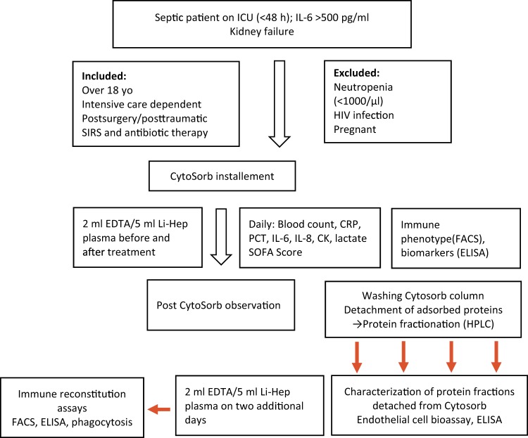
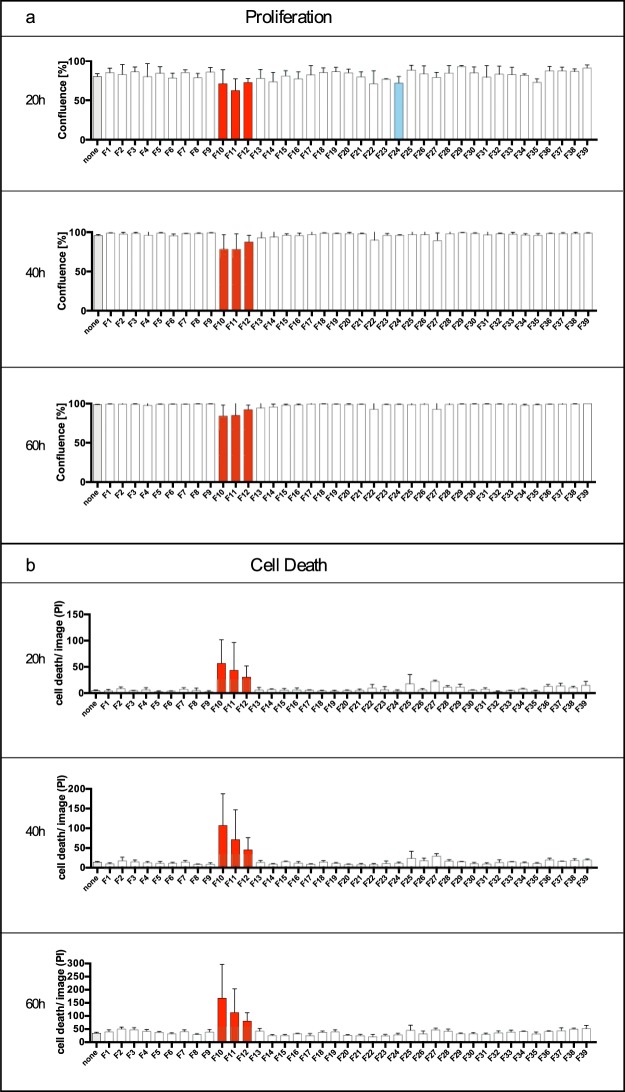
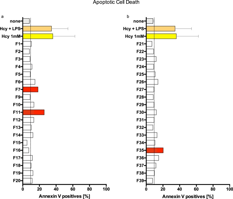
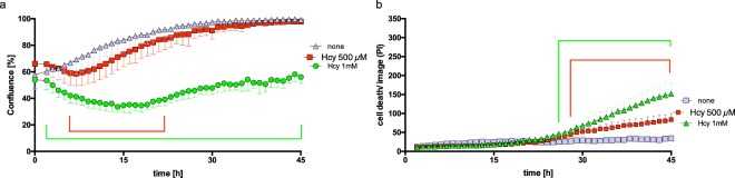
Hemoadsorption devices are used to treat septic shock by adsorbing inflammatory cytokines and as yet incompletely defined danger and pathogen associated molecular patterns. In an ideal case, hemoadsorption results in immediate recovery of microvascular endothelial cells' (mEC) function and rapid recovery from catecholamine-dependency and septic shock. We here tested a single device, which consists of polystyrene-divinylbenzene core particles of 450 μm diameter with a high affinity for hydrophobic compounds. The current study aimed at the proof of concept that endothelial-specific damage mediators are adsorbed and can be recovered from hemoadsorption devices. Because of excellent clinical experience, we tested protein fractions released from a hemoadsorber in a novel endothelial bioassay. Video-based, long-term imaging of mEC proliferation and cell death were evaluated and combined with apoptosis and ATP measurements. Out of a total of 39 fractions recovered from column fractionation, we identified 3 fractions that caused i) inhibition of mEC proliferation, ii) increased cell death and iii) induction of apoptosis in mEC. When adding these 3 fractions to mEC, their ATP contents were reduced. These fractions contained proteins of approximately 15 kDa, and high amounts of nucleic acid, which was at least in part oxidized. The efficacy for endothelial cell damage prevention by hemoadsorption can be addressed by a novel endothelial bioassay and long-term video observation procedures. Protein fractionation of the hemoadsorption devices used is feasible to study and define endothelial damage ligands on a molecular level. The results suggest a significant effect by circulating nucleic acids - bound to an as yet undefined protein, which may constitute a major danger-associated molecular pattern (DAMP) in the exacerbation of inflammation when patients experience septic shock. Hemoadsorption devices may thus limit endothelial damage, through the binding of nucleic acid-bearing aggregates and thus contribute to improved endothelial barrier function.


Conflict of interest statement

The authors declare no competing interests.

Figures

Figure 1
Figure 1
Flowchart of our Working Procedure. This schematic describes the process instructions for hemoadsorption on ICU and Clinical Study to identify mediators removed by hemoadsorption and documentation of a potential improvement of the patient.
Figure 2
Figure 2
Pattern of CytoSorb protein fractions on microvascular endothelial cells (mEC) growth and cell death. mEC were cultured in the absence (none) and presence of 1:10 diluted protein fractions (F) for 60 hours in total, with individual measurements at every hour: Confluency was determined by optical densities using the IncuCyteZOOM. Protein fractions resulting in significantly diminished confluence rates are shown as red (F10–F12) and blue (F24) columns following two-way ANOVA (multiple comparison) (a). Protein fractions resulting in cell death of cultured mEC were determined by adding 2 ng/ml propidium iodide to the culture medium; counting of red fluorescent nuclei was accomplished by IncuCyteZOOM videomicroscopy and red object count quantification. Fractions leading to significantly increased cell death are shown by a red column (b). Relevant time points of 20 hours, 40 hours and 60 hours are shown of the continuous observation. Significances were determined by two-way ANOVA (multiple comparison). All values are given as means and standard deviation (of triplicate cultures) for statistically relevant protein fractions. Images were obtained with the IncuCyteZOOM using a 20× objective, phase contrast and fluorescence detection (565–605 nm excitation, 625–705 nm emission).
Figure 3
Figure 3
Modulation of Annexin V expression by microvascular endothelial cells (mEC). mEC were cultivated in petri dishes (2 × 105 cells per dish) and incubated for 24 hours with reduced L-homocysteine (Hcy, 1 mM) with or without lipopolysaccharide (LPS, 10 µg/ml) or with CytoSorb-derived protein fractions at a final dilution of 1:10. Cells were detached using trypsin and stained with Annexin V and propidium iodide (PI). Flow cytometry was performed at a final cell count of 5 × 104 per assay. Annexin V-positive cells are shown. For control (none) and positive control (Hcy, Hcy + LPS) means of four replicates and standard deviation (SD) are displayed, SD of the control is shown (dotted line). Protein fractions F7 and F11 induced apoptosis, with F11 resulting in more than three times more Annexin V-positive cells when compared to the control (25.80% vs. 7.99%) (a). Protein fraction F35 also induced apoptosis in more than twice as many cells as in the control (20.30% vs. 7.99%) (b).
Figure 4
Figure 4
Microvascular endothelial cells (mEC) are sensitive to the known toxic agent L-homocysteine (Hcy). Endothelial cell growth in the absence (control) and presence of 0.5 mM or 1 mM reduced Hcy was monitored for 45 hours (x-axis). For fluorescent detection of cell death, PI was used (0.05 mg/ml). Confluency was measured by optical analysis with the IncuCyteZOOM software using a confluence mask and given as percent (y-axis). Adding 1 mM reduced Hcy resulted in decreased (from 0 hours 53.9% to 14 hours 33.8%) confluency and inhibited cell growth to 56% of the control (45 hours). Compared to the control, we found a dose-dependent decrease in mEC confluency, by 8% at 7 hours for 0.5 mM and by 20% at 14 hours for 1 mM Hcy. The difference in confluency compared to control was statistically significant between 1 hour and 45 hours (green bracket) (multiple comparison two-way ANOVA, p < 0.0001) (a). Cell death is displayed as the red object count (dead cells)/image (y-axis)). Reduced Hcy at 1 mM showed significantly more cell death/image compared to the control at 26 hours to 45 hours (green bracket) (multiple comparison two-way ANOVA, p < 0.0001). Values are given as means, while standard deviation (triplicates) is shown for the statistically relevant protein fractions (b).
Figure 5
Figure 5
Morphology of microvascular endothelial cells (mEC) C2-6 in the absence and presence of homocysteine. Scanning electron micrograph of mEC cultured in the absence (a,b) and in the presence of L-homocysteine for 24 hours (c,d). Note the impaired cell-to-cell contact formation and high density of stress-associated microvilli in Hcy-treated mEC.
Figure 6
Figure 6
ATP contents in microvascular endothelial cells (mEC) after 6 and 20 hours of incubation with CytoSorb-derived protein fractions. ATP contents in the absence (none) and presence of protein fractions (F, diluted 1:10 v/v) were recorded for 6 hours (left column; a–c) and 20 hours (right column; d–f). Cells were lysed after 6 and 20 hours and intracellular ATP contents were determined as relative luminescent units (RLU) (ATPCellTiter Glo, Promega). After 6 hours of incubation, some protein fractions showed significantly higher ATP levels when compared to the control: F2 (p < 0.05); F5 (p < 0.01); F6 (p < 0.05); F7 (p < 0.01); F8 (p < 0.05); F39 (p < 0.01) (Dunnett´s multiple comparison test); whereas protein fraction F25 diminished ATP contents (p < 0.05) when compared to the control (ac). After 20 hours of incubation, the following protein fractions decreased ATP contents in mEC: F8 (p < 0.05); F9 (p < 0.01); F11 (p < 0.01); F14 (p < 0.05) (Dunnett´s multiple comparison test) (d–f). Values are given as means and standard deviation of triplicates. The results were representative for two independent experiments. All fractions with a different trend of either upregulation or downmodulation of ATP contents at 6 hours and 20 hours of incubation were labeled by blue border lined bars and fractions consistently impairing ATP contents were labeled by red-bordered columns (F10–F12, F24).
Figure 7
Figure 7
Characterization of CytoSorb-derived protein fractions. Albumin contents (ng/ml, y-axis) in given fractions (x-axis) using a high-sensitive ELISA (Immulite1000®). Fractions F1–F18 and F26–F39 were below the detection limit (4 µg/ml). The highest amount of albumin was found in F20 (>6000 µg/ml). (a) Amounts of deoxyribonucleic acids (DNA) were determined by spectrophotometry at 260/280 nm (NanoDrop). The highest amounts of DNA were found in F13 (3915.6 ng/ml) (b) the insert shows the contents of oxidized DNA (8-hydroxy-2′-deoxyguanosine from DNA, 8-hydroxyguanosine from RNA and 8-hydroxyguanine from DNA or RNA); results were obtained by ELISA (Cayman Chemicals.com); the highest concentration was found in F11 (40.97 pg/ml). Advanced glycation end products (AGEs) were quantified by ELISA (mybiosource.com); the highest amount was found in F14 (0.95 µg/ml); means of duplicates’ analyses are shown. (c) Red-filled bars were fractions which consistently influenced endothelial viability, ATP contents, and apoptosis.

References

    1. Singer M, et al. The Third International Consensus Definitions for Sepsis and Septic Shock (Sepsis-3) JAMA. 2016;315:801–810. doi: 10.1001/jama.2016.0287. - DOI - PMC - PubMed
    1. Ince C, et al. The Endothelium in Sepsis. Shock. 2016;45:259–270. doi: 10.1097/SHK.0000000000000473. - DOI - PMC - PubMed
    1. Lipcsey M, et al. Abdominal Septic Shock - Endotoxin Adsorption Treatment (ASSET) - endotoxin removal in abdominal and urogenital septic shock with the Alteco(R) LPS Adsorber: study protocol for a double-blinded, randomized placebo-controlled trial. Trials. 2016;17:587–016. doi: 10.1186/s13063-016-1723-4. - DOI - PMC - PubMed
    1. Cruz DN, et al. Early use of polymyxin B hemoperfusion in abdominal septic shock: the EUPHAS randomized controlled trial. JAMA. 2009;301:2445–2452. doi: 10.1001/jama.2009.856. - DOI - PubMed
    1. Reinhart K, Bauer M, Riedemann NC, Hartog CS. New approaches to sepsis: molecular diagnostics and biomarkers. Clin. Microbiol. Rev. 2012;25:609–634. doi: 10.1128/CMR.00016-12. - DOI - PMC - PubMed

Publication types