. 2019 Oct 10;9(1):14650.
doi: 10.1038/s41598-019-51195-9.

Keratin 19 regulates cell cycle pathway and sensitivity of breast cancer cells to CDK inhibitors

Affiliations

Keratin 19 regulates cell cycle pathway and sensitivity of breast cancer cells to CDK inhibitors

Pooja Sharma et al. Sci Rep. .

Abstract

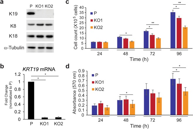
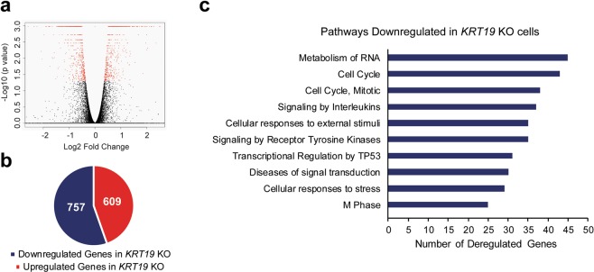
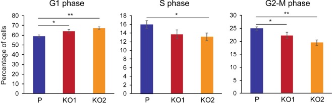
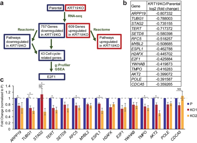
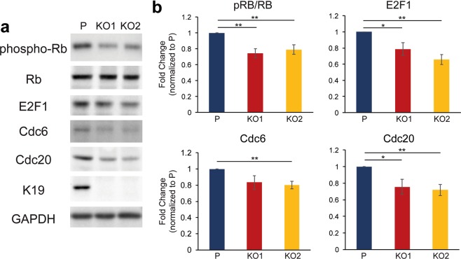
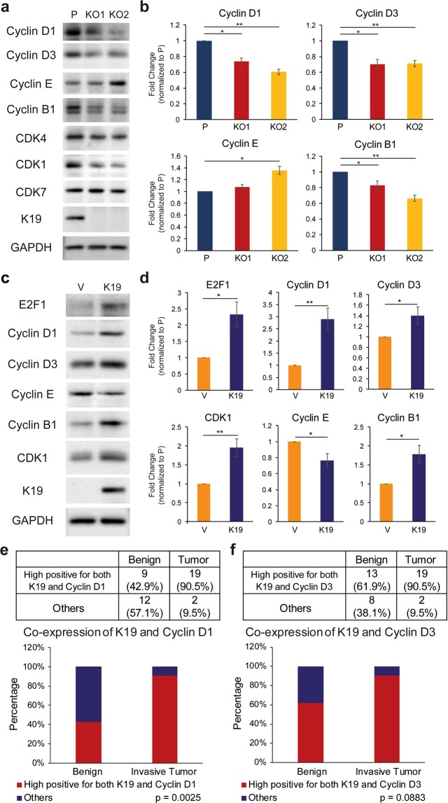
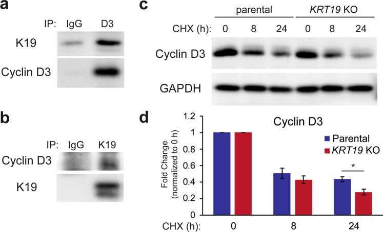
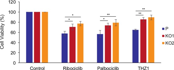
Keratin 19 (K19) belongs to the keratin family of proteins, which maintains structural integrity of epithelia. In cancer, K19 is highly expressed in several types where it serves as a diagnostic marker. Despite the positive correlation between higher expression of K19 in tumor and worse patient survival, the role of K19 in breast cancer remains unclear. Therefore, we ablated K19 expression in MCF7 breast cancer cells and found that K19 was required for cell proliferation. Transcriptome analyses of KRT19 knockout cells identified defects in cell cycle progression and levels of target genes of E2F1, a key transcriptional factor for the transition into S phase. Furthermore, proper levels of cyclin dependent kinases (CDKs) and cyclins, including D-type cyclins critical for E2F1 activation, were dependent on K19 expression, and K19-cyclin D co-expression was observed in human breast cancer tissues. Importantly, K19 interacts with cyclin D3, and a loss of K19 resulted in decreased protein stability of cyclin D3 and sensitivity of cells towards CDK inhibitor-induced cell death. Overall, these findings reveal a novel function of K19 in the regulation of cell cycle program and suggest that K19 may be used to predict the efficacy of CDK inhibitors for treatments of breast cancer.


Conflict of interest statement

The authors declare no competing interests.

Figures

Figure 1
Figure 1
Keratin 19 knockout cells exhibit reduced proliferation rate. (a) Whole cell lysates of parental (P) control and two different clones (KO1 and KO2) of KRT19 KO cell lines were harvested, and immunoblotting was performed with antibodies against the indicated proteins. (b) qRT-PCR performed showing mRNA levels of K19 in indicated cells. *p < 1 × 10−7. Data from three experimental repeats normalized to the parental control are shown as mean ± SEM. Proliferation of cells were assessed by (c) counting cells and (d) performing MTT assay and measuring the absorbance at 570 nm each day following cell plating. Data from at least four experimental repeats are shown as mean ± SEM. Differences are not statistically significant unless denoted by *p < 0.05; **p < 1 × 10−4.
Figure 2
Figure 2
Decreased expression of transcripts related to cell cycle-related pathways in KRT19 KO cells. (a) A volcano plot for all genes or transcript isoforms using R Studio package. The data for all transcripts were plotted as log2 fold change versus the −log10 of the adjusted p-value. Transcripts selected as significantly different are based on FDR-adjusted p value < 0.05 and highlighted as red dots. (b) Number of differentially expressed genes or transcript isoforms (FDR-adjusted p value < 0.05) observed in KRT19 KO versus parental cells. (c) Pathways related to genes downregulated in KRT19 KO cells.
Figure 3
Figure 3
KRT19 KO cells show defects in cell cycle progression. Cells were synchronized in serum-starved media for 48 h before treating them with 10% serum-containing media for 24 h. Cells were then fixed, nuclei stained with propidium iodide and analyzed using flow cytometry. Percentage of cells from four experimental repeats are shown as mean ± SEM. Differences are not statistically significant unless denoted by *p < 0.05; **p < 0.0005.
Figure 4
Figure 4
Identification of E2F1 as a regulator of K19-dependent gene expression. (a) A schematic of bioinformatical analyses performed to identify E2F1 as a transcriptional regulator of a subset of genes downregulated in KRT19 KO cells. (b) 15 genes identified to have putative E2F1 regulatory motifs through g:Profiler and GSEA are shown with log2 fold from RNA-sequencing results in Supplementary Table S2. (c) Levels of 15 E2F1-regulated targets in parental and KRT19 KO cells were verified using qRT-PCR. Data from at least five experimental repeats normalized to that of the parental control are shown as mean ± SEM. All differences between parental and KRT19 KO cells are statistically significant with p < 0.05 unless denoted by *p < 0.01 or NS, not significant.
Figure 5
Figure 5
Decreased levels of Rb phosphorylation and E2F1 in KRT19 KO cells. (a) Whole cell lysates of P, KO1, and KO2 cells were harvested, and immunoblotting was performed with antibodies against the indicated proteins. (b) Signal intensities of bands from (a) were quantified and normalized to those of the GAPDH loading control. Data from at least three experimental repeats normalized to that of the parental control are shown as mean ± SEM. Differences are not statistically significant unless denoted by *p < 0.05; **p < 0.003.
Figure 6
Figure 6
K19-dependent expression of cyclins and CDKs. (a) Whole cell lysates of P, KO1, and KO2 cells were harvested, and immunoblotting was performed with antibodies against the indicated proteins. (b) Signal intensities of cyclins from (a) were quantified and normalized to those of the GAPDH loading control. Data from at least three experimental repeats normalized to that of the parental control are shown as mean ± SEM. Differences are not statistically significant unless denoted by *p < 0.05; **p < 0.001. (c) Whole cell lysates of KRT19 KO cells expressing vector (V) or K19 (K19) were harvested, and immunoblotting was performed with antibodies against the indicated proteins. (d) Signal intensities of bands from (c) were quantified and normalized to those of GAPDH loading control. Data from at least three experimental repeats normalized to that of the parental control are shown as mean ± SEM. Differences are not statistically significant unless denoted by *p < 0.05; **p < 0.001. Tissue sections from 21 differerent breast cancer patients with most aggressive tumors were immunostained for (e) K19 and cyclin D1 or (f) K19 and cyclin D3. The immunoreactivity of cells in both the invasive tumor and adjacent benign epithelium in each case were scored and categorized as shown in Supplementary Tables S6 and 7. Those that were strongly positive in both K19 and cyclin D1 or cyclin D3 were separated from cases that were not (others).
Figure 7
Figure 7
K19 physically interacts with cyclin D3 and is required for cyclin D3 stability. Co-IP was performed with (a) anti-cyclin D3 (D3) or (b) anti-K19 antibody with IgG as a control. Immunoblotting was performed with antibodies against the indicated proteins. (c) Whole cell lysates of P and KRT19 KO2 (KO) cells treated with either 20 ng/µl of cycloheximide for indicated time points or DMSO vehicle control (0 h) were harvested, and immunoblotting was performed with antibodies against the indicated proteins. (d) Signal intensities of cyclin D3 from (c) were quantified and normalized to the GAPDH loading control. Data from at least three experimental repeats normalized to their respective vehicle control are shown as mean ± SEM. Differences are not statistically significant unless denoted by *p < 0.05.
Figure 8
Figure 8
KRT19 KO cells exhibit decreased sensitivity towards CDK inhibitors. (a) MTT assays were performed on P, KO1, and KO2 cells after cells were grown for 72 h in the presence of 250 nM ribociclib, 500 nM palbociclib, 25 nM THZ1, or DMSO control. The absorbance at 570 nm of drug-treated cells was normalized to that of its respective DMSO control to calculate cell viability. Data from at least three experimental repeats are shown as mean ± SEM. Differences are not statistically significant unless denoted by *p < 0.05; **p < 0.002.

References

    1. Pan X, Hobbs RP, Coulombe PA. The expanding significance of keratin intermediate filaments in normal and diseased epithelia. Curr. Opin. Cell Biol. 2013;25:47–56. doi: 10.1016/j.ceb.2012.10.018. - DOI - PMC - PubMed
    1. Kim S, Coulombe PA. Intermediate filament scaffolds fulfill mechanical, organizational, and signaling functions in the cytoplasm. Genes Dev. 2007;21:1581–1597. doi: 10.1101/gad.1552107. - DOI - PubMed
    1. Chung BM, Rotty JD, Coulombe PA. Networking galore: intermediate filaments and cell migration. Curr. Opin. Cell Biol. 2013;25:600–612. doi: 10.1016/j.ceb.2013.06.008. - DOI - PMC - PubMed
    1. Moll R, Franke WW, Schiller DL, Geiger B, Krepler R. The catalog of human cytokeratins: Patterns of expression in normal epithelia, tumors and cultured cells. Cell. 1982;31:11–24. doi: 10.1016/0092-8674(82)90400-7. - DOI - PubMed
    1. Moll R, Divo M, Langbein L. The human keratins: biology and pathology. Histochem. Cell Biol. 2008;129:705–733. doi: 10.1007/s00418-008-0435-6. - DOI - PMC - PubMed

Publication types

MeSH terms