Review
. 2020 Jan;20(1):26-41.
doi: 10.1038/s41568-019-0205-x. Epub 2019 Oct 10.

The blood-brain barrier and blood-tumour barrier in brain tumours and metastases

Affiliations
Review

The blood-brain barrier and blood-tumour barrier in brain tumours and metastases

Costas D Arvanitis et al. Nat Rev Cancer. 2020 Jan.

Abstract

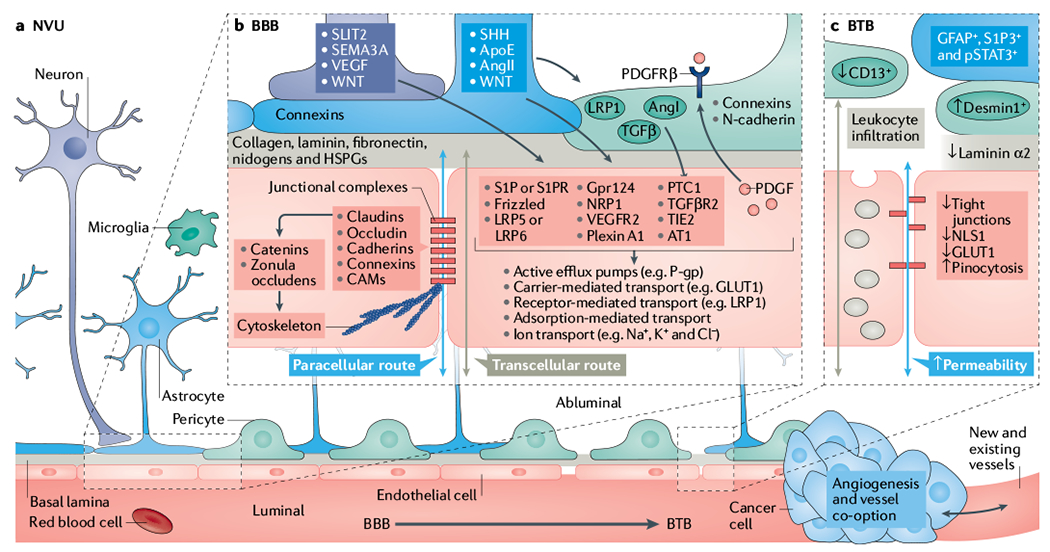
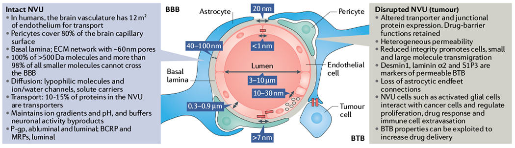
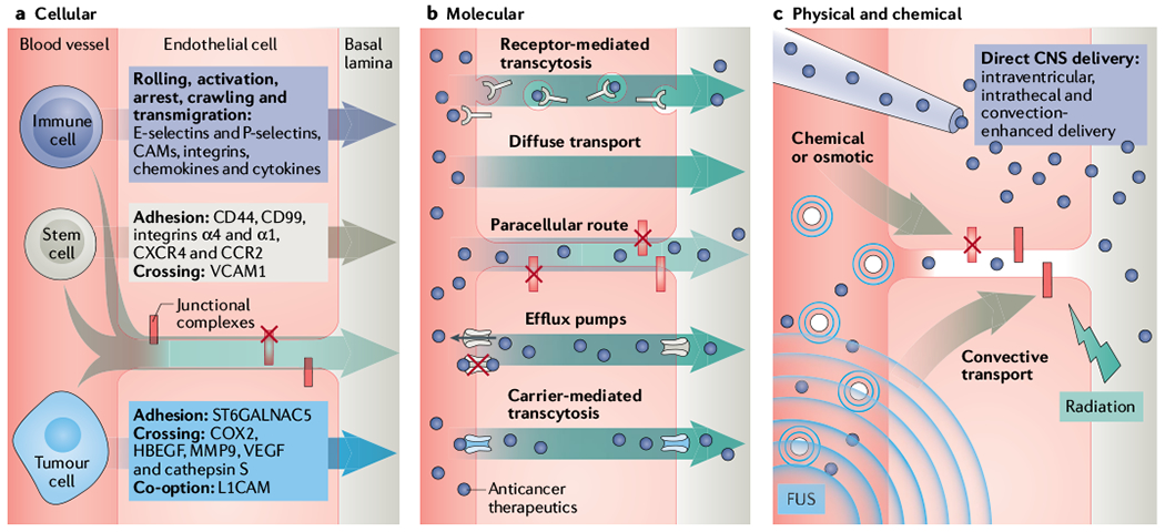
For a blood-borne cancer therapeutic agent to be effective, it must cross the blood vessel wall to reach cancer cells in adequate quantities, and it must overcome the resistance conferred by the local microenvironment around cancer cells. The brain microenvironment can thwart the effectiveness of drugs against primary brain tumours as well as brain metastases. In this Review, we highlight the cellular and molecular components of the blood-brain barrier (BBB), a specialized neurovascular unit evolved to maintain brain homeostasis. Tumours are known to compromise the integrity of the BBB, resulting in a vasculature known as the blood-tumour barrier (BTB), which is highly heterogeneous and characterized by numerous distinct features, including non-uniform permeability and active efflux of molecules. We discuss the challenges posed by the BBB and BTB for drug delivery, how multiple cell types dictate BBB function and the role of the BTB in disease progression and treatment. Finally, we highlight emerging molecular, cellular and physical strategies to improve drug delivery across the BBB and BTB and discuss their impact on improving conventional as well as emerging treatments, such as immune checkpoint inhibitors and engineered T cells. A deeper understanding of the BBB and BTB through the application of single-cell sequencing and imaging techniques, and the development of biomarkers of BBB integrity along with systems biology approaches, should enable new personalized treatment strategies for primary brain malignancies and brain metastases.


Figures

Fig. 1 |
Fig. 1 |. Overview of the neurovascular unit in healthy and tumour-bearing brains.
a | Schematic representation of capillaries in the neurovascular unit (NVU) with the intact blood–brain barrier (BBB, bottom left) and the disrupted blood–tumour barrier (BTB, bottom right) in the neuroparenchyma. BBB development and permeability is dictated by signalling and structural mechanisms that are regulated by multiple cells within the NVU. These mechanisms control both paracellular and transcellular routes and, ultimately, vessel permeability in the central nervous system (CNS). b | Schematic representation of notable cellular and molecular components that regulate the development, maturation and function of endothelial cells (ECs) (red) and the NVU (refer to main text for relevant references). Neuronal (purple) and non-neuronal cells regulate the expression of transport and tight junction proteins in ECs, which in turn may ‘loosen’ or ‘tighten’ the BBB. Here, we depict examples of key signalling pathways connecting astrocytes (blue), pericytes (green) and neurons to ECs. Together or individually, these pathways will alter transcellular transport by changing the expression of transporters and the paracellular route by disrupting junctional protein complexes. Of note, ECs reciprocally regulate components of the NVU. For example, EC-secreted transforming growth factor-β (TGFβ) can activate cognate receptor on pericytes. During development and maturation, glial cells, pericytes and neurons regulate EC behaviour via multiple ligands and receptors, which in turn activate downstream signalling cascades (for example, Frizzled, G protein-coupled receptor 124 (GPR124), β-catenin, GLI, PI3K, SRC and the p38 MAPK) that dictate expression of junctional and transcytosis proteins and control CNS homeostasis. For example, astrocytes directly modulate NVU demands such as water content in the neuroparenchymal space via the major water channel protein aquaporin 4 (AQP4), regulate immune cell and cancer cell infiltration via specific chemokine and cytokine production, regulate BBB permeability and integrity in part by angiotensin (AngI and AngII), apolipoprotein E (ApoE) and retinoic acid, and regulate pericyte distribution. c | In the BTB, NVU integrity and endothelial permeability is compromised due to disruption of the NVU, including displacement of astrocytes (blue) and pericytes (green), neurovascular decoupling, altered pericyte populations and changes in EC tight junctions and transcytosis mechanisms. Additional vascular-related phenotypes such as hypoxia, oedema, angiogenesis and tumour-vessel co-option can influence the NVU in brain tumours. Although BBB features remain present during tumour development, in particular at the cancer–neuroparenchyma edge, the BTB displays increased and heterogeneous permeability. Tumour progression leads to BTB structural changes including neuronal death, astrocyte endfeet displacement (from primary and metastatic cancer cells) and heterogenous pericyte and astrocyte subpopulations, all of which can reduce the barrier functions of the CNS endothelium. Intracellular vesicular transport is represented by grey-coloured vesicles in the schematic. AT1, angiotensin II receptor type 1; CAM, cell adhesion molecule; GLUT1, glucose transporter 1; HSPG, heparan sulfate proteoglycan; LRP, low-density lipoprotein receptor-related protein; NLS1, sodium-dependent lysophosphatidylcholine symporter 1; NRP1, neuropilin receptor 1; PDGF, platelet-derived growth factor; PDGFRβ, platelet-derived growth factor receptor β; P-gp, P-glycoprotein; PTC1, protein patched homologue 1; S1P, sphingosine-1-phosphate; S1PR, sphingosine-1-phosphate receptor; SEMA3A, semaphorin 3A; SHH, sonic hedgehog; SLIT2, Slit homologue 2 protein; TGFβR2, transforming growth factor-β receptor 2; TIE2, tyrosine kinase with Ig and EGF homology domains 2; VEGF, vascular endothelial growth factor; VEGFR2, vascular endothelial growth factor receptor 2.
Fig. 2 |
Fig. 2 |. Physical and chemical properties of the BBB.
The blood–brain barrier (BBB) structure displays unique physical properties that tightly regulate molecular and cellular flow in the neuroparenchyma. The brain barrier’s importance is evident from its functional conservation across organisms, from fruit flies to humans. As the BBB develops and matures, endothelial cell fenestrations decrease, and the subsequent appearance of tight junctions is followed by a reduction in transcytosis. Centre panel: cross-section of a central nervous system (CNS) capillary depicting estimated distances and spaces within the BBB, which circulating drugs are required to overcome to permeate the brain parenchyma. Left panel: in the non-diseased brain, the neurovascular unit (NVU) includes an intact BBB that displays multiple characteristics that limit drug permeability into the CNS. Right panel: during tumour progression, stroma–cancer cell interactions in the brain tumour microenvironment dictate vessel permeability and cancer cell proliferation. The blood–tumour barrier (BTB) characteristics listed here contribute to the heterogeneous permeability observed in the disrupted NVU. BCRP, breast cancer resistance protein; ECM, extracellular matrix; MRP, multidrug resistance protein; P-gp, P-glycoprotein; S1P3, sphingosine 1-phosphate 3.
Fig. 3 |
Fig. 3 |. Improving drug delivery through the BBB/BTB.
Schematic presentation of key molecular, cellular and physical mechanisms and systems to overcome the blood–brain barrier and blood–tumour barrier (BBB/BTB). a | The BBB prevents cellular extravasation into the neuroparenchyma unless compromised by circulating cells equipped with necessary ‘brain-tropic’ molecular components, including soluble and membrane-bound proteins, to disturb the BBB integrity. Immune cell extravasation into the central nervous system (CNS) occurs by the following steps: rolling, activation, arrest, crawling, transmigration. The transcellular route is preferred when the BBB is intact, whereas the paracellular route is preferred when there is reduced tight junction integrity (red X = disrupted junction) and formation of intercellular gaps. Transmigration across the BBB is mediated by actin-containing protrusive structures and occurs on the timescale of minutes. Stem cells that have been engineered to contain anticancer cargo localize to sites of neuroinflammation and display coordinated rolling and adhesion behaviour, and transcellular and paracellular transmigration. In contrast to immune cells, mesenchymal stem cell (MSC) transmigration does not involve substantial lateral crawling. Stem cells migrate by the paracellular or transcellular route through discrete gaps or pores in the CNS endothelium. Stem cell transmigration is mediated by membrane blebbing and occurs on the timescale of hours. In contrast, circulating metastatic cancer cells that accumulate in the CNS capillary bed must express specific proteins in order to adhere and breach the BBB. Metastatic cells express proteases that disrupt junctional complexes. Although preclinical studies show that this process may occur within days, the time course of symptomatic brain metastasis in patients varies greatly (from months to years after metastatic dissemination of primary tumour). b | Several molecular strategies are employed to hijack or bypass barriers posed by the neurovascular unit (NVU) (described in main text and TABLE 1). Anticancer therapeutics (purple circles) can be designed with low affinity to efflux pumps or may hijack carriers or receptor-mediated transcytosis mechanisms. Alternatively, inhibitors of efflux pump or junctional complexes can be used to limit the clearance of drugs from the tumours. c | Direct delivery into the neuroparenchyma and physical disruption of the BBB/BTB. Specifically, focused ultrasound (FUS, indicated by the waves) with microbubbles (concentric small blue circles), radiation, osmotic, direct and convective-mediated delivery of therapeutics into the CNS is depicted. CAM, cell adhesion molecule; CCR2, CC-chemokine receptor 2; COX2, cyclooxygenase 2; CXCR4, CXC-chemokine receptor 4; HBECF, proheparin-binding ECF-like growth factor; L1CAM, L1 cell adhesion molecule; ST6GALNAC5, a 2,6-sialyltransferase; MMP9, matrix metalloproteinase 9; VCAM1, vascular cell adhesion protein 1; VECF, vascular endothelial growth factor.

References

    1. O’Brown NM, Pfau SJ & Gu C Bridging barriers: a comparative look at the blood–brain barrier across organisms. Genes Dev 32, 466–478 (2018). - PMC - PubMed
    2. This review describes the conservation and variation of BBB function across organisms.

    1. Abbott NJ Blood–brain barrier structure and function and the challenges for CNS drug delivery. J. Inherit. Metab. Dis 36, 437–449 (2013). - PubMed
    1. Hobbs SK et al. Regulation of transport pathways in tumor vessels: role of tumor type and microenvironment. Proc. Natl Acad. Sci. USA 95, 4607–4612 (1998). - PMC - PubMed
    2. This study presents the first systematic examination of the role of the brain versus the subcutaneous microenvironment on permeability of tumour vessels.

    1. Monsky WL et al. Role of host microenvironment in angiogenesis and microvascular functions in human breast cancer xenografts: mammary fat pad versus cranial tumors. Clin. Cancer Res 8, 1008–1013 (2002). - PubMed
    2. This study demonstrates the differential response of blood vessels in tumours growing in the brain versus in the subcutaneous microenvironment.

    1. Pitz MW, Desai A, Grossman SA & Blakeley JO Tissue concentration of systemically administered antineoplastic agents in human brain tumors. J. Neurooncol 104, 629–638 (2011). - PMC - PubMed

Publication types

Substances