

Anatomy of Landsmeer Ligaments-Redefined
- PMID: 31602135
- PMCID: PMC6785317
- DOI: 10.1055/s-0039-1695802
Anatomy of Landsmeer Ligaments-Redefined
Abstract
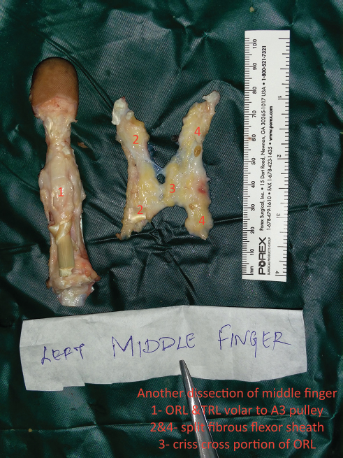
Introduction Landsmeer ligaments play a significant role in synchronizing the movements of the two distal phalanges of the fingers. However, there is considerable controversy in descriptions of its anatomy, function, presence, and clinical applications. Aim To ascertain and redefine the detailed anatomy of Oblique Retinacular (ORL) and Transverse Retinacular ligaments (TRL) and their applied features. Materials and Methods Anatomical dissection study was conducted in 100 cadaveric fingers in 42 cadavers (28 fresh specimens and remaining preserved specimens) under loupe magnification. The whole dorsal digital expansion with attached fibrous flexor sheath was dissected and specimen was examined after thorough saline wash. The dimensions, course, attachment, and configuration were noted in each specimen. The statistical mean was obtained for thickness of the ligaments. The measurements were made using a caliper at the level of the mid proximal phalanx, volar to the proximal interphalangeal (PIP) joint, and dorsal to the distal interphalangeal (DIP) joint. Results By anatomical dissection we have found the following: • The ORL was deep to the TRL. • The ORL had got a check rein effect at the PIP joint, in such a way that extension of the PIP joint causes extension of the DIP joint. • The ORL criss-crossed volar to the A3 pulley of fibrous flexor sheath and formed a good hammock for the PIP joint. This criss-crossing anatomical feature was found in all dissected fingers as an additional normal anatomical feature complementing the classical description of Landsmeer. • Variations in configuration of the Landsmeer ligaments were observed among various fingers. • The Landsmeer ligament was never absent as reported by several studies. Conclusion Contrary to several studies, the ORL was omnipresent in all dissected fingers with considerable variations in dimensions. Complementing the classical description of Landsmeer, we found that there was an additional normal criss-cross anatomical feature of the ORL in all fingers volar to A3 pulley and deep to the TRL. Also, the TRL was present in all the fingers.
Keywords: criss-cross hammock of the oblique retinacular ligament; oblique retinacular ligament; pleomorphism of the Landsmeer ligament; redefinition of Landsmeer ligament anatomy; transverse retinacular ligament.
Conflict of interest statement
Figures


References
-
- el-Gammal T A, Steyers C M, Blair W F, Maynard J A. Anatomy of the oblique retinacular ligament of the index finger. J Hand Surg Am. 1993;18(04):717–721. - PubMed
-
- Landsmeer J M. The anatomy of the dorsal aponeurosis of the human finger and its functional significance. Anat Rec. 1949;104(01):31–44. - PubMed
-
- Shrewsbury M M, Johnson R K. A systematic study of the oblique retinacular ligament of the human finger: its structure and function. J Hand Surg Am. 1977;2(03):194–199. - PubMed
-
- Shrewsbury M M, Johnson R K. Ligaments of the distal interphalangeal joint and the mallet position. J Hand Surg Am. 1980;5(03):214–216. - PubMed
LinkOut - more resources
Full Text Sources