. 2019 Dec 1;317(6):H1258-H1271.
doi: 10.1152/ajpheart.00478.2019. Epub 2019 Oct 11.

BDNF downregulates β-adrenergic receptor-mediated hypotensive mechanisms in the paraventricular nucleus of the hypothalamus

Affiliations

BDNF downregulates β-adrenergic receptor-mediated hypotensive mechanisms in the paraventricular nucleus of the hypothalamus

Daniella Thorsdottir et al. Am J Physiol Heart Circ Physiol. .

Abstract

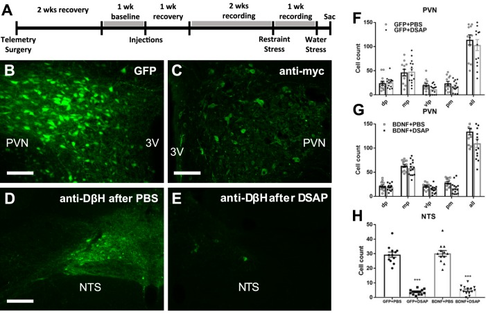
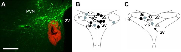
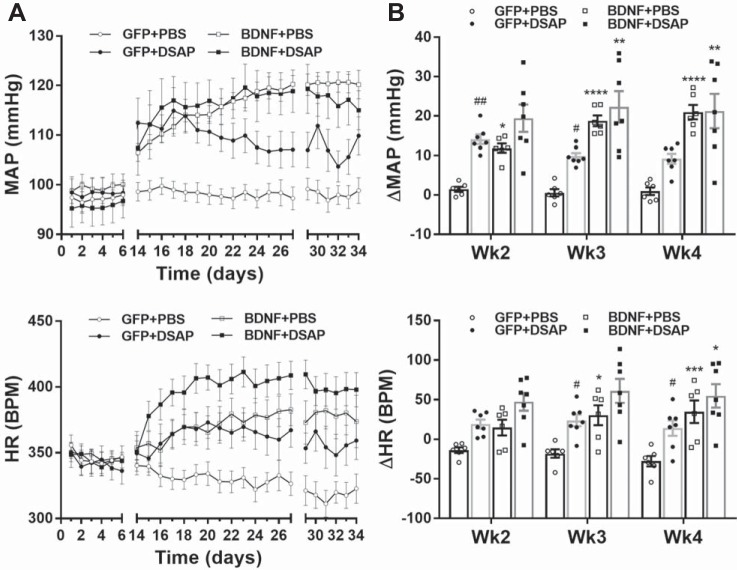
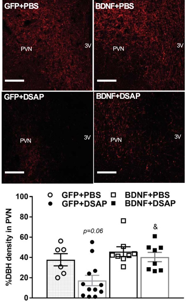
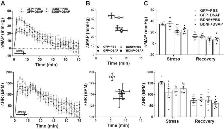
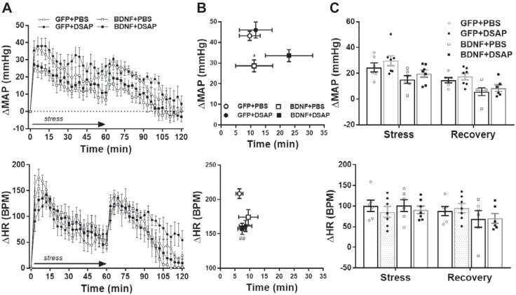
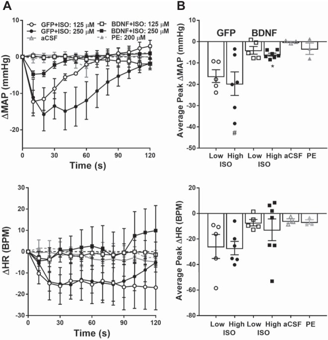
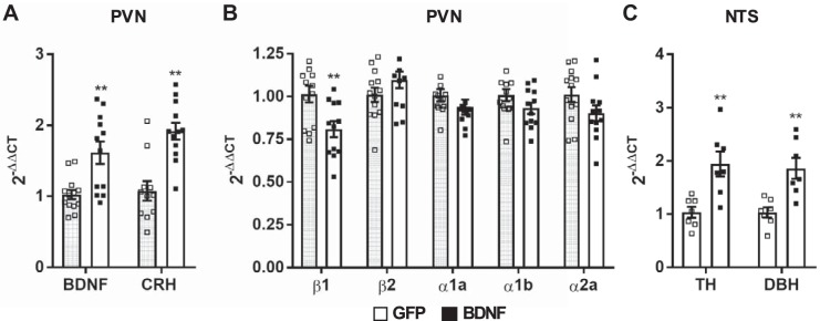
Brain-derived neurotrophic factor (BDNF) is upregulated in the paraventricular nucleus of the hypothalamus (PVN) in response to hypertensive stimuli such as stress and hyperosmolality, and BDNF acting in the PVN plays a key role in elevating sympathetic activity and blood pressure. However, downstream mechanisms mediating these effects remain unclear. We tested the hypothesis that BDNF increases blood pressure, in part by diminishing inhibitory hypotensive input from nucleus of the solitary tract (NTS) catecholaminergic neurons projecting to the PVN. Male Sprague-Dawley rats received bilateral PVN injections of viral vectors expressing either green fluorescent protein (GFP) or BDNF and bilateral NTS injections of vehicle or anti-dopamine-β-hydroxylase-conjugated saporin (DSAP), a neurotoxin that selectively lesions noradrenergic and adrenergic neurons. BDNF overexpression in the PVN without NTS lesioning significantly increased mean arterial pressure (MAP) in awake animals by 18.7 ± 1.8 mmHg. DSAP treatment also increased MAP in the GFP group, by 9.8 ± 3.2 mmHg, but failed to affect MAP in the BDNF group, indicating a BDNF-induced loss of NTS catecholaminergic hypotensive effects. In addition, in α-chloralose-urethane-anesthetized rats, hypotensive responses to PVN injections of the β-adrenergic agonist isoprenaline were significantly attenuated by BDNF overexpression, whereas PVN injections of phenylephrine had no effect on blood pressure. BDNF treatment was also found to significantly reduce β1-adrenergic receptor mRNA expression in the PVN, whereas expression of other adrenergic receptors was unaffected. In summary, increased BDNF expression in the PVN elevates blood pressure, in part by downregulating β-receptor signaling and diminishing hypotensive catecholaminergic input from the NTS to the PVN.NEW & NOTEWORTHY We have shown that BDNF, a key hypothalamic regulator of blood pressure, disrupts catecholaminergic signaling between the NTS and the PVN by reducing the responsiveness of PVN neurons to inhibitory hypotensive β-adrenergic input from the NTS. This may be occurring partly via BDNF-mediated downregulation of β1-adrenergic receptor expression in the PVN and results in an increase in blood pressure.

Keywords: blood pressure; brain-derived neurotrophic factor; catecholamine; hypothalamus; β-adrenergic receptors.


Conflict of interest statement

No conflicts of interest, financial or otherwise, are declared by the authors.

Figures

Fig. 1.
Fig. 1.
A: timeline of experiment 1. B and C: representative fluorescent images of coronal brain sections ~1.8 mm posterior from bregma showing PVN expression of GFP and BDNFmyc, respectively. BDNFmyc expression was detected with an anti-myc tag antibody and immunofluorescence; scale bars = 100 µm. D and E: representative fluorescence images showing DβH expression in the NTS in coronal brain sections at the level of the calamus scriptorius following PBS and DSAP injections, respectively; scale bars, 100 µm. F and G: number of GFP- and myc-positive cells was assessed in subnuclei of the PVN; dorsal parvocellular nuclei (dp), medial parvocellular nuclei (mp), ventrolateral parvocellular nuclei (vlp), and posterior magnocellular nuclei (pm) following AAV2-GFP or AAV2-BDNFmyc injections in the PVN and PBS or DSAP injections in the NTS. DSAP had no significant effect on vector mediated gene transduction in GFP or BDNF rats. GFP+PBS (n = 12), GFP+DSAP (n = 14), BDNF+PBS (n = 12), BDNF+DSAP (n = 14); n refers to number of PVN sides. H: number of DβH-positive neurons in the NTS following AAV2-GFP or AAV2-BDNFmyc injections in the PVN and PBS or DSAP injections in the NTS. DSAP significantly reduced the number of DβH-positive neurons in both the GFP and BDNF groups. 3V, third ventricle; PVN, paraventricular nucleus; NTS, nucleus of the solitary tract; GFP, green fluorescent protein; PBS, phosphate-buffered saline; BDNF, brain-derived neurotrophic factor; BDNFmyc, myc epitope-tagged BDNF; DβH, dopamine β-hydroxylase; DSAP, anti-dopamine-β-hydroxylase-conjugated saporin; AAV2, adeno-associated viral vector 2. ***P < 0.001 for DSAP (1-way ANOVA).
Fig. 2.
Fig. 2.
A: representative fluorescent image of a coronal brain section ~1.8 mm posterior to bregma showing PVN expression of GFP and the red fluorescent microbead solution that was mixed with injected drug solution to verify injection sites; scale bars, 250 µm. B and C: diagrams of the PVN ~1.8 mm and ~1.9 mm posterior to bregma showing locations of isoprenaline injections in rats previously injected with AAV2-GFP (gray circles) or AAV2-BDNFmyc (filled circles), and phenylephrine (open circles) and aCSF (open triangles) injections in untreated rats. 3V, third ventricle; lm, lateral magnocellular nuclei; dp, dorsal parvocellular nuclei; mp, medial parvocellular nuclei; vlp, ventrolateral parvocellular nuclei; PVN, paraventricular nucleus; GFP, green fluorescent protein; BDNF, brain-derived neurotrophic factor; BDNFmyc, myc epitope-tagged BDNF; AAV2, adeno-associated viral vector 2; aCSF, artificial cerebrospinal fluid.
Fig. 3.
Fig. 3.
Radiotelemetric recordings of daytime mean arterial pressure (MAP) and heart rate [HR, in beats/min (bpm)]. Radiotelemetric parameters were recorded for 6 days before brain injections and during weeks 2–4 after brain injections. Recording was turned off during the postsurgical recovery phase (between days 6 and 14), and data from day 28 are omitted because animals were subjected to water stress on that day. Since viral vector and DSAP treatments had similar effects on daytime and nighttime parameters, only daytime parameters are shown (nighttime data are summarized in Table 1). A: daytime MAP (top) and HR (bottom) in GFP+PBS (n = 6), GFP+DSAP (n = 7), BDNF+PBS (n = 6), and BDNF+DSAP (n = 7) rats. B: average changes in daytime MAP (top) and HR (bottom) during weeks 2, 3, and 4 from pretreatment baseline period. PVN, paraventricular nucleus; NTS, nucleus of the solitary tract; GFP, green fluorescent protein; PBS, phosphate-buffered saline; BDNF, brain-derived neurotrophic factor; DSAP, anti-dopamine-β-hydroxylase-conjugated saporin. Results represent means ± SE. Two-way repeated-measures ANOVA on weekly averages indicated significant treatment effect for MAP and HR (P < 0.001), significant time effect for HR (P < 0.05), and significant treatment × time interaction for both MAP and HR (P < 0.01). Post hoc analysis indicated *P < 0.05 for GFP+PBS vs. BDNF+PBS, **P < 0.01 for GFP+DSAP vs. BDNF+DSAP, ***P < 0.001 for GFP+PBS vs. BDNF+PBS, ****P < 0.0001 for GFP+PBS vs. BDNF+PBS; #P < 0.05 for GFP+PBS vs. GFP+DSAP, ##P < 0.01 for GFP+PBS vs. GFP+DSAP.
Fig. 4.
Fig. 4.
DβH-positive vesicles in the PVN of rats previously injected with AAV2-GFP or AAV2-BDNFmyc in the PVN and with PBS or DSAP in the NTS. Top: representative confocal images of DβH expression within the PVN in all experimental groups as detected with an anti-DβH antibody and immunofluorescence; scale bars, 100 µm. Bottom: maximum percent volumetric density of DβH-positive vesicles within the PVN, averaged for each experimental group; n refers to number of PVN sides [GFP+PBS (n = 6), GFP+DSAP (n = 12), BDNF+PBS (n = 8), BDNF+DSAP (n = 8)]. 3V, third ventricle; PVN, paraventricular nucleus; NTS, nucleus of the solitary tract; GFP, green fluorescent protein; PBS, phosphate-buffered saline; BDNF, brain-derived neurotrophic factor; DBH, dopamine β-hydroxylase; DSAP, anti-dopamine-β-hydroxylase-conjugated saporin; AAV2, adeno-associated viral vector 2. Results represent means ± SE. P = 0.06 for GFP+PBS vs. GFP+DSAP; &P < 0.05 for GFP+DSAP vs. BDNF+DSAP (1-way ANOVA).
Fig. 5.
Fig. 5.
Radiotelemetric recordings of mean arterial pressure (MAP) and heart rate (HR) during acute water stress and poststress recovery in GFP+PBS (n = 6), GFP+DSAP (n = 7), BDNF+PBS (n = 6) and BDNF+DSAP (n = 7) rats. A: MAP (top) and HR (bottom) traces with 3-min moving average during stress (15 min, indicated by arrow) and during poststress recovery (60 min). B: amplitude and delay of peak MAP and HR responses. C: MAP and HR increases averaged during stress and poststress recovery. Prestress MAP and HR were 94 ± 3 mmHg and 308 ± 11 beats/min (bpm) in the GFP+PBS group, 101 ± 4 mmHg and 327 ± 9 beats/min in the GFP+DSAP group, 121 ± 4 mmHg and 366 ± 19 beats/min in the BDNF+PBS group, and 113 ± 5 mmHg and 378 ± 11 beats/min in the BDNF+DSAP group. GFP, green fluorescent protein; PBS, phosphate-buffered saline; BDNF, brain-derived neurotrophic factor; DSAP, anti-dopamine-β-hydroxylase-conjugated saporin. Results represent means ± SE. **P < 0.01 for GFP+PBS vs. BDNF+PBS (1-way ANOVA).
Fig. 6.
Fig. 6.
Radiotelemetric recordings of mean arterial pressure (MAP) and heart rate (HR) during acute restraint stress and poststress recovery in GFP+PBS (n = 6), GFP+DSAP (n = 7), BDNF+PBS (n = 6), and BDNF+DSAP (n = 7) rats. A: MAP (top) and HR (bottom) traces with 3-min moving average during stress (60 min, indicated by arrow) and during poststress recovery (60 min). B: amplitude and delay of peak MAP and HR responses. C: MAP and HR increases averaged during stress and poststress recovery. Prestress MAP and HR were 92 ± 3 mmHg and 311 ± 11 beats/min (bpm) in the GFP+PBS group, 101 ± 4 mmHg and 351 ± 14 beats/min in the GFP+DSAP group, 120 ± 2 mmHg and 372 ± 18 beats/min in the BDNF+PBS group, and 114 ± 3 mmHg and 380 ± 13 beats/min in the BDNF+DSAP group. GFP, green fluorescent protein; PBS, phosphate-buffered saline; BDNF, brain-derived neurotrophic factor; DSAP, anti-dopamine-β-hydroxylase-conjugated saporin. Results are represented as means ± SE. *P < 0.05, for GFP+PBS vs. BDNF+PBS, ##P < 0.01, GFP+PBS vs. GFP+DSAP (1-way ANOVA).
Fig. 7.
Fig. 7.
Changes in mean arterial pressure (MAP; top) and heart rate (HR; bottom) in response to PVN injections of adrenergic receptor agonists and vehicle in anesthetized rats previously treated with AAV2-GFP (n = 6) or AAV2-BDNFmyc (n = 7). A: MAP and HR obtained with 10-s moving average over 120 s following isoprenaline (ISO) injection in GFP and BDNFmyc treated rats, and phenylephrine (PE; n = 3) or aCSF, artificial cerebrospinal fluid (aCSF; n = 3) injections in untreated rats. B: average peak MAP and HR responses to ISO, PE, and aCSF injections. Baseline MAP and HR were 89 ± 6 mmHg and 351 ± 15 beats/min (bpm) in GFP and 108 ± 4 mmHg and 398 ± 15 beats/min in BDNF rats, and 105 ± 3 mmHg and 353 ± 7 beats/min in untreated rats. PVN, paraventricular nucleus; GFP, green fluorescent protein; BDNF, brain-derived neurotrophic factor; BDNFmyc, myc epitope-tagged BDNF; AAV2, adeno-associated viral vector 2. Results represent means ± SE. Statistical analysis was done on peak changes in MAP and HR. #P < 0.05 for 250 µM ISO vs. aCSF, *P < 0.05, for GFP vs. BDNF (1-way ANOVA).
Fig. 8.
Fig. 8.
Expression of BDNF, CRH, and adrenergic receptor mRNA in the PVN, and TH and DβH mRNA in the NTS of rats previously injected with AAV2-GFP (n = 6) or AAV2-BDNFmyc (n = 6) in the PVN. A: significant increases in BDNF and CRH mRNA expressions verify successful vector mediated BDNF transduction and activation of BDNF-mediated signaling mechanisms. B: expressions of α1a-, α1b-, and α2a- and β1- and β2-adrenergic receptor mRNAs in GFP and BDNF rats. C: expressions of TH and DβH mRNAs in the NTS of AAV2-GFP (n = 7) and AAV2-BDNFmyc (n = 7) rats. PVN, paraventricular nucleus; NTS, nucleus of the solitary tract; GFP, green fluorescent protein; BDNF, brain-derived neurotrophic factor; BDNFmyc, myc epitope-tagged BDNF; TH, tyrosine hydroxylase; DBH, dopamine β-hydroxylase; AAV2, adeno-associated viral vector 2; CRH, corticotropin-releasing hormone. Results are expressed as 2−ΔΔCT (where CT is threshold cycle) normalized to the control group and presented as means ± SE. **P < 0.01 GFP vs. BDNF (unpaired t test).

Similar articles

Cited by

References

    1. Aliaga E, Arancibia S, Givalois L, Tapia-Arancibia L. Osmotic stress increases brain-derived neurotrophic factor messenger RNA expression in the hypothalamic supraoptic nucleus with differential regulation of its transcripts. Relation to arginine-vasopressin content. Neuroscience 112: 841–850, 2002. doi:10.1016/S0306-4522(02)00128-8. - DOI - PubMed
    1. Andresen MC, Kunze DL. Nucleus tractus solitarius--gateway to neural circulatory control. Annu Rev Physiol 56: 93–116, 1994. doi:10.1146/annurev.ph.56.030194.000521. - DOI - PubMed
    1. Becker BK, Tian C, Zucker IH, Wang HJ. Influence of brain-derived neurotrophic factor-tyrosine receptor kinase B signalling in the nucleus tractus solitarius on baroreflex sensitivity in rats with chronic heart failure. J Physiol 594: 5711–5725, 2016. doi:10.1113/JP272318. - DOI - PMC - PubMed
    1. Becker BK, Wang HJ, Tian C, Zucker IH. BDNF contributes to angiotensin II-mediated reductions in peak voltage-gated K+ current in cultured CATH.a cells. Physiol Rep 3: e12598, 2015. doi:10.14814/phy2.12598. - DOI - PMC - PubMed
    1. Bhatia V, Rarick KR, Stauss HM. Effect of the data sampling rate on accuracy of indices for heart rate and blood pressure variability and baroreflex function in resting rats and mice. Physiol Meas 31: 1185–1201, 2010. doi:10.1088/0967-3334/31/9/009. - DOI - PubMed

Publication types

MeSH terms