. 2019 Oct 15;51(4):709-723.e6.
doi: 10.1016/j.immuni.2019.09.005. Epub 2019 Oct 8.

Calcitonin Gene-Related Peptide Negatively Regulates Alarmin-Driven Type 2 Innate Lymphoid Cell Responses

Affiliations

Calcitonin Gene-Related Peptide Negatively Regulates Alarmin-Driven Type 2 Innate Lymphoid Cell Responses

Antonia Wallrapp et al. Immunity. .

Abstract

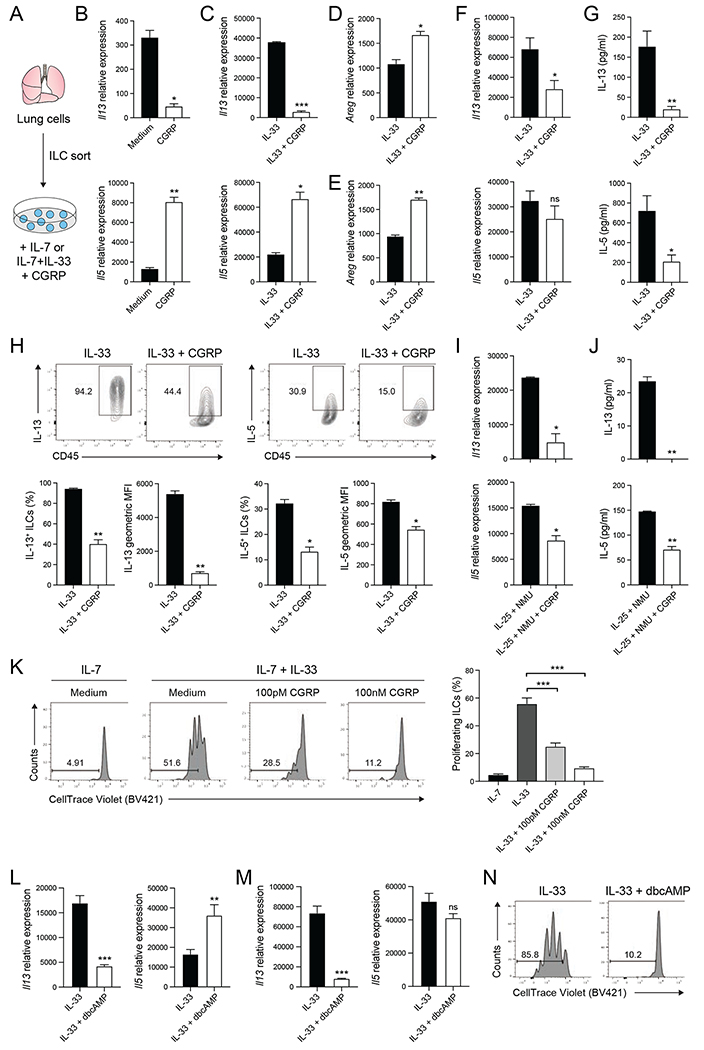
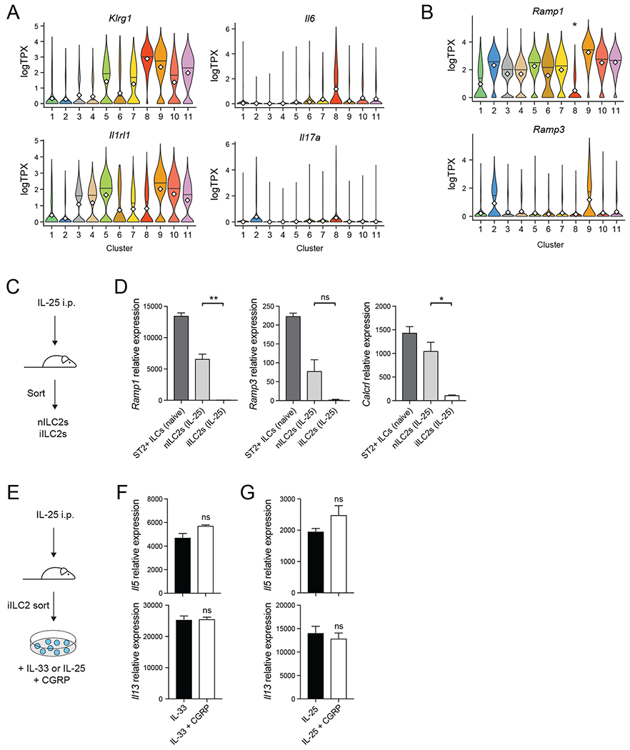
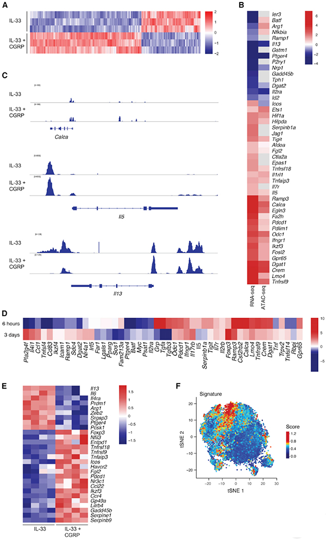
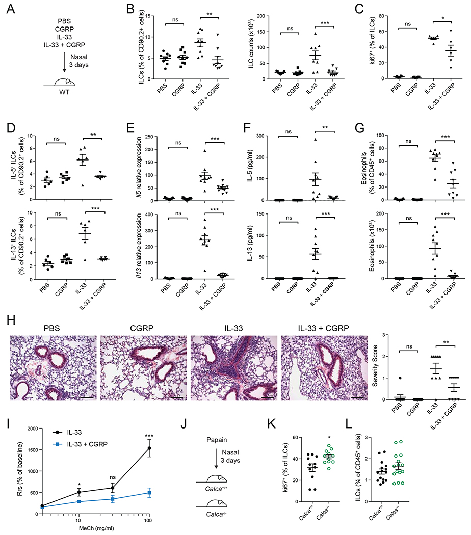
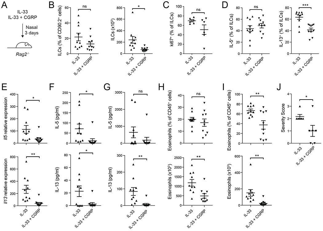
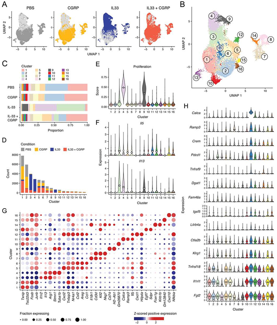
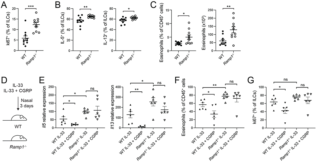
Neuroimmune interactions have emerged as critical modulators of allergic inflammation, and type 2 innate lymphoid cells (ILC2s) are an important cell type for mediating these interactions. Here, we show that ILC2s expressed both the neuropeptide calcitonin gene-related peptide (CGRP) and its receptor. CGRP potently inhibited alarmin-driven type 2 cytokine production and proliferation by lung ILC2s both in vitro and in vivo. CGRP induced marked changes in ILC2 expression programs in vivo and in vitro, attenuating alarmin-driven proliferative and effector responses. A distinct subset of ILCs scored highly for a CGRP-specific gene signature after in vivo alarmin stimulation, suggesting CGRP regulated this response. Finally, we observed increased ILC2 proliferation and type 2 cytokine production as well as exaggerated responses to alarmins in mice lacking the CGRP receptor. Together, these data indicate that endogenous CGRP is a critical negative regulator of ILC2 responses in vivo.

Keywords: CGRP; Ramp1; airway inflammation; allergic inflammation; neuro-immune interaction; neuropeptides; type 2 innate lymphoid cells.


Conflict of interest statement

Declaration of Interests

A.R. is an SAB member of ThermoFisher Scientific, Neogene Therapeutics, and Syros Pharmaceuticals. A.R. and V.KK. are cofounders of and equity holders in Celsius Therapeutics. A.W., P.R.B., S.J.R., A.R. and V.K.K. are co-inventors on US provisional patent application no. 62/667,381 and A.R. and R.J.X. are co-inventors on US provisional patent application no. 62/818,404, which are both filed by The Broad Institute relating to CGRP signaling modulation. All other authors have no competing financial interests.

Figures

Figure 1.
Figure 1.. CGRP inhibits type 2 cytokine expression and proliferation of ILC2s in vitro
(A) Lungs ILCs were isolated from C57BL/6J mice by fluorescence activated cell sorting (FACS) and cultured with IL-7 or IL-7+IL-33 with medium or CGRP. (B-C) Il13 and Il5 expression in ILC2s cultured for 6 hours, determined by qPCR. Data points are technical replicates (n = 2). Data are representative of two independent experiments. (D-E) Areg expression in ILC2s cultured for either 6 hours (D) or 3 days (E), determined by qPCR. Data points are technical replicates (n = 2). Data are representative of two (D) or three (E) independent experiments. (F) Il13 and Il5 expression in ILCs cultured for 3 days, determined by qPCR. Data points are averages of technical replicates from four independent experiments. (G) IL-13 and IL-5 concentration in 3 day ILC culture supernatants, determined by LegendPlex. Data points are averages of technical replicates from four independent experiments. (H) Expression of IL-13 and IL-5 in ILCs cultured for 3 days, analyzed by flow cytometry. Representative flow cytometry plots, frequency and geometric mean fluorescence intensity (MFI) of IL-5 and IL-13 are shown. Data points are technical replicates (n = 2). Data are representative of two independent experiments. (I) Il13 and Il5 expression in ILCs cultured with IL-25+NMU or IL-25+NMU+CGRP for 3 days, determined by qPCR. Data points are technical replicates from one experiment. Data are representative of three independent experiments. (J) IL-13 and IL-5 concentration in 3 day ILC culture supernatants, determined by LegendPlex. Data points are technical replicates from one experiment. Data are representative of three independent experiments. (K) CellTrace Violet-labeled ILCs cultured for 3 days, analyzed by flow cytometry. Histograms (left) show CellTrace Violet dye dilution. Graph (right) shows the frequency of proliferating ILCs from 2-3 independent experiments. (L) Il13 and Il5 expression in ILC2s cultured for 6 hours, determined by qPCR. Data are pooled from three independent experiments with two technical replicates each. (M) Il13 and Il5 expression in ILCs cultured for 3 days, determined by qPCR. Data are pooled from three independent experiments with two technical replicates each. (N) Cell Trace Violet-labeled ILCs were cultured for 3 days and proliferation was analyzed by flow cytometry. Data are representative of two independent experiments. Data shown as the mean ± s.e.m. *P <0.05; **P <0.01; ***P <0.001; ns, not significant; by either unpaired t test (B-J, L, M) or unpaired ANOVA (K). Please see also Figure S2.
Figure 2.
Figure 2.. Inflammatory ILC2s express less Ramp1 and do not respond to CGRP
(A-B) Expression of the indicated genes in ILCs by cluster (as shown in Figure S1D) as determined by scRNA-seq. Violin plots show expression of Klrg1, Il6 Il1rl1, and Il17a (A), or Ramp1 and Ramp3 (B) by cluster (x axis). *P<4.3x10−45, logistic regression, Wald test. (C) IL-25 was given intraperitoneally for three consecutive days. Natural ILC2s (ST2+ KLRG1-ILCs; nILC2s) and inflammatory ILC2s (ST2-KLRG1+ ILCs; iILC2s) were then isolated. (D) Ramp1, Ramp3 and Calcrl expression in different ILC subsets, determined by qPCR. Data points are technical replicates from one experiment. Data are representative of two independent experiments. (E) iILC2s were cultured in vitro with IL-33, IL-33+CGRP, IL-25 or IL-25+CGRP for 6 hours. (F-G) Il5 and Il13 expression in iILC2s, determined by qPCR. Data points are technical replicates from one experiment. Data are representative of two independent experiments. For panels (D,F,G) data are shown as the mean ± s.e.m. *P <0.05; **P <0.01; ns, not significant; by either unpaired ANOVA (D) or unpaired t test (F,G). Please see also Figure S3.
Figure 3.
Figure 3.. CGRP modulates ILC activation and induces a regulatory gene module
(A) ILC2s cultured for 6 hours with IL-33 or IL-33+CGRP were profiled by ATAC-seq. Accessible regions are mapped as z-scores of normalized peak counts taken across columns (n = 3). (B) ILC2s cultured for 6 hours with IL-33 or IL-33+CGRP were analyzed by ATAC-seq and bulk RNA-sequencing (RNA-seq). Heatmap of significant fold changes (>1.5, color) in expression (left column) of genes (rows) in ILC2s cultured with IL-33+CGRP versus IL-33 (FDR-adjusted P<0.05, generalized linear regression, Wald test). Fold changes in chromatin accessibility (right) are shown for the ATAC-seq peak corresponding to the respective gene by GREAT. When multiple peaks were assigned to a gene, the peak with the most significant change between ILC2s cultured with IL-33+CGRP versus IL-33 was shown (FDR-adjusted P<0.05, Wald test). (C) Chromatin accessibility in ILC2s treated with IL-33 (upper track of each set) or IL-33+CGRP (lower track of each set) is shown for the Calca, Il5 and Il13 loci. (D) ILCs cultured for 3 days with IL-33 or IL-33+CGRP were analyzed by bulk RNA-seq. Heatmap of significant fold changes (>1.5, color) in expression of genes (rows) in ILC2s cultured with IL-33+CGRP versus IL-33 for 6 hours (upper row, cultured as described in B) or 3 days (lower row). (FDR-adjusted P<0.05, generalized linear regression, Wald test.) (E) Heatmap of expression (color), z-scored by row, of selected pro-inflammatory and regulatory genes (rows) that exhibit a significant fold change (>1.5) in expression of at least 1.5 in ILCs cultured with IL-33 (left) versus IL-33+CGRP (right) for 3 days. Each column represents one replicate. (FDR-adjusted P<0.05, generalized linear regression, Wald test.) (F) tSNE plot of published single-cell RNA-seq data, as in Figure S1C,D, shows ILCs (dots) isolated from PBS-, IL-25- or IL-33-treated mice, colored by score of CGRP signature. Please see also Figure S4 and S5.
Figure 4.
Figure 4.. CGRP dampens IL-33-induced airway inflammation
(A) PBS, CGRP, IL-33 or IL-33+CGRP were administered nasally daily for 3 days. (B) Frequency (left) and number (right) of lung ILCs, determined by flow cytometry. (C) Frequency of Ki67+ lung ILCs, determined by flow cytometry. (D) Frequency of IL-5+ and IL-13+ lung ILCs, determined by flow cytometry. (E) Il5 and Il13 expression in lung tissue, determined by qPCR. (F) IL-5 and IL-13 concentration in BALF, determined by LegendPlex. (G) Eosinophil frequency and number in BALF, determined by flow cytometry. (H) Representative H&E stained lung sections from mice treated with PBS, CGRP, IL-33 or IL-33+CGRP (left). Graph (right) shows severity score for individual mice (n = 9) from three independent experiments. Scale bars represent 100μm. (I) Airway resistance was assessed in IL-33− or IL-33+CGRP-treated mice in response to methacholine challenge. Data points represent the mean of individual mice from two independent experiments (IL-33, n = 9; IL-33+CGRP, n = 9). (J) Calca+/+ and Calca−/− mice were treated nasally with papain for three days. (K) Frequency of Ki67+ lung ILCs, determined by flow cytometry. (L) Lung ILC frequency, determined by flow cytometry. Data points are individual mice pooled from three independent experiments (n = 9) in panels B and E-G and from two independent experiments (n = 6) in panels C and D. Data points are individual mice pooled from three independent experiments (n = 11) in panel K and from four independent experiments (n=15) in panel L. Data are shown as the mean ± s.e.m. *P <0.05; **P <0.01; ***P <0.001; ns, not significant; by either unpaired ANOVA (B-H) or unpaired t test (I, K, L). Please see also Figure S6.
Figure 5.
Figure 5.. CGRP negatively regulates ILC2 responses in vivo independent of adaptive immunity
(A) Nasal administration of IL-33 or IL-33+CGRP to Rag2−/− mice for three consecutive days. (B) Frequency (left) and number (right) of lung ILCs, determined by flow cytometry. (C) Frequency of Ki67+ ILCs, analyzed by flow cytometry. (D) Frequency of IL-5+ and IL-13+ lung ILCs, determined by flow cytometry. (E) Il5 and Il13 expression in lung tissue, determined by qPCR. (F-G) IL-5 and IL-13 concentration in lung tissue (F) and BALF (G), determined by LegendPlex. (H-I) Frequency and number of eosinophils in lung tissue (H) and BALF (I), determined by flow cytometry. (J) Lung sections were scored for disease severity in a blinded manner. Graph shows severity score for individual mice (n = 6) from two independent experiments. Data points represent individual mice (n = 10) pooled from three independent experiments (B,D-I) or individual mice (n=6) from two independent experiments (C, J). Data are shown as the mean ± s.e.m. *P <0.05; **P <0.01; ***P <0.001; ns, not significant; by unpaired t test.
Figure 6.
Figure 6.. CGRP inhibits proliferative and inflammatory response of ILC2s to IL-33
(A,B) Uniform manifold approximation and projection (UMAP) embedding of 31,053 cells (dots) isolated from PBS-, CGRP-, IL-33-, or IL-33+CGRP-treated mice, colored by treatment condition (A) or by cluster (B). (C) Bar plot of proportions (x axis) of cells from each cluster (color) per condition (y axis). (D) Bar plot of counts (y axis) of cells from each treatment condition (color) per cluster (x axis). (E) ILC2s were scored for expression of proliferation signature genes. Violin plots show distributions of proliferation signature scores (y axis) for each cluster (x axis). (F) Violin plots show distributions of Il5 and Il13 expression (y axis, white diamond indicates mean) for each cluster (x axis). (G) Dot plot visualizes the mean expression (color), z-scored by column, in positively expressing cells of selected significantly differentially expressed genes (x axis, Methods), per cluster (y axis). Dot size represents the proportion of cells in a cluster that express the gene. (H) Violin plots show distributions of expression (y axis, white diamond indicates mean) in each cluster (x axis, color) of selected genes (rows) that are significantly differentially expressed in Cluster 11 (blue) compared with all other cells (Methods). Please see also Figure S7.
Figure 7.
Figure 7.. Increased type 2 immunity in Ramp1−/− mice
(A-G) WT mice were purchased from Jackson Laboratories and co-housed with Ramp1−/− mice for at least one week. (A-C) Naive WT and Ramp1−/− mice. (A) Frequency of Ki67+ lung ILCs, analyzed by flow cytometry. (B) Frequency of IL-5+ and IL-13+ lung ILCs, determined by flow cytometry. (C) Lung eosinophil frequency and number, analyzed by flow cytometry. (D-G) IL-33 or IL-33+CGRP were administered nasally to WT mice or Ramp1−/− mice for 3 days. (E) Il5 and I113 expression in lung tissue, determined by qPCR. (F) BALF eosinophil frequency, analyzed by flow cytometry. (G) Frequency of Ki67+ lung ILCs, determined by flow cytometry. Data points are individual mice pooled from two independent experiments (n = 10, panels A-C or n= 6, panels E-G). Data are shown as the mean ± s.e.m. *P <0.05; **P <0.01; ***P <0.001; ns, not significant; by unpaired t-test.

Comment in

References

    1. Aoki-Nagase T, Nagase T, Oh-Hashi Y, Shindo T, Kurihara Y, Yamaguchi Y, Yamamoto H, Tomita T, Ohga E, Nagai R, et al. (2002). Attenuation of antigen-induced airway hyperresponsiveness in CGRP-deficient mice. Am J Physiol Lung Cell Mol Physiol 283, L963–970. - PubMed
    1. Baliu-Pique M, Jusek G, and Holzmann B (2014). Neuroimmunological communication via CGRP promotes the development of a regulatory phenotype in TLR4-stimulated macrophages. European journal of immunology 44, 3708–3716. - PubMed
    1. Baral P, Umans BD, Li L, Wallrapp A, Bist M, Kirschbaum T, Wei Y, Zhou Y, Kuchroo VK, Burkett PR, et al. (2018). Nociceptor sensory neurons suppress neutrophil and gammadelta T cell responses in bacterial lung infections and lethal pneumonia. Nature medicine 24, 417–426. - PMC - PubMed
    1. Barnig C, Cernadas M, Dutile S, Liu X, Perrella MA, Kazani S, Wechsler ME, Israel E, and Levy BD (2013). Lipoxin A4 regulates natural killer cell and type 2 innate lymphoid cell activation in asthma. Sci Transl Med 5, 174ra126. - PMC - PubMed
    1. Bettelli E, Carrier Y, Gao W, Korn T, Strom TB, Oukka M, Weiner HL, and Kuchroo VK (2006). Reciprocal developmental pathways for the generation of pathogenic effector TH17 and regulatory T cells. Nature 441, 235–238. - PubMed

Publication types

MeSH terms