Comparative Study
. 2019 Dec;213(4):1545-1563.
doi: 10.1534/genetics.119.302749. Epub 2019 Oct 11.

Comparative Genomics and Transcriptomics To Analyze Fruiting Body Development in Filamentous Ascomycetes

Affiliations
Comparative Study

Comparative Genomics and Transcriptomics To Analyze Fruiting Body Development in Filamentous Ascomycetes

Ramona Lütkenhaus et al. Genetics. 2019 Dec.

Abstract

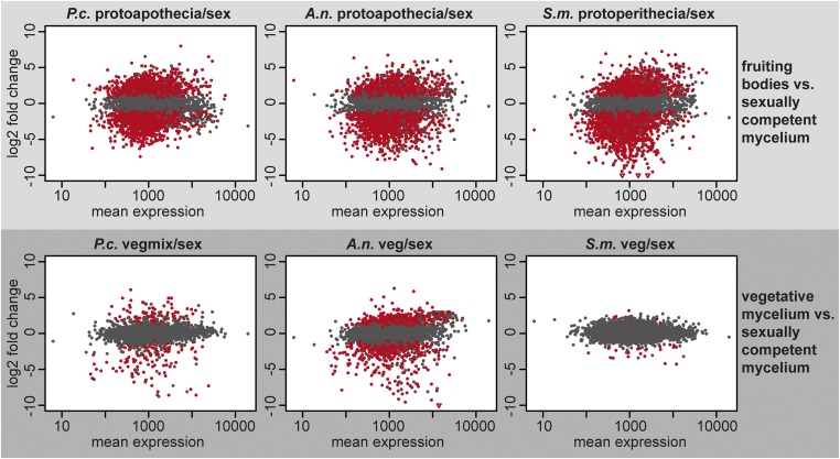
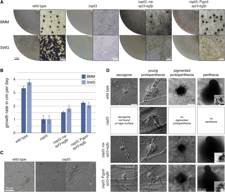
Many filamentous ascomycetes develop three-dimensional fruiting bodies for production and dispersal of sexual spores. Fruiting bodies are among the most complex structures differentiated by ascomycetes; however, the molecular mechanisms underlying this process are insufficiently understood. Previous comparative transcriptomics analyses of fruiting body development in different ascomycetes suggested that there might be a core set of genes that are transcriptionally regulated in a similar manner across species. Conserved patterns of gene expression can be indicative of functional relevance, and therefore such a set of genes might constitute promising candidates for functional analyses. In this study, we have sequenced the genome of the Pezizomycete Ascodesmis nigricans, and performed comparative transcriptomics of developing fruiting bodies of this fungus, the Pezizomycete Pyronema confluens, and the Sordariomycete Sordaria macrospora With only 27 Mb, the A. nigricans genome is the smallest Pezizomycete genome sequenced to date. Comparative transcriptomics indicated that gene expression patterns in developing fruiting bodies of the three species are more similar to each other than to nonsexual hyphae of the same species. An analysis of 83 genes that are upregulated only during fruiting body development in all three species revealed 23 genes encoding proteins with predicted roles in vesicle transport, the endomembrane system, or transport across membranes, and 13 genes encoding proteins with predicted roles in chromatin organization or the regulation of gene expression. Among four genes chosen for functional analysis by deletion in S. macrospora, three were shown to be involved in fruiting body formation, including two predicted chromatin modifier genes.

Keywords: Ascodesmis nigricans; Pyronema confluens; Sordaria macrospora; comparative transcriptomics; fruiting body development.


Figures

Figure 1
Figure 1
Life cycle of A. nigricans under continuous illumination and laboratory conditions. Strain CBS 389.68 was grown on microscopic slides with RFA medium (with 0.8% agar) for 1–6 days in constant light. After 1 day, a mycelium of septated hyphae is formed. After 2 days, apothecia initials can be observed that contain swollen young asci after 3 days (arrows). Immature hyaline spores can be observed within asci after 4 days. Spores become pigmented during maturation after 5 days. Mature spores are released from eight-spore asci after 6 days. Development of mycelium and apothecia is the same in constant darkness (Figure S1). Bar for all images, 20 µm.
Figure 2
Figure 2
Species tree of 20 fungal species based on phylome reconstruction. The species tree was built based on 143 single-copy, widespread genes (see Materials and Methods for details). All nodes are maximally supported by 100% bootstrap. The scale bar gives substitutions per site.
Figure 3
Figure 3
Comparison of the mating type loci of A. nigricans and P. confluens. Orthologs of two genes that are linked to MAT1-2-1 in P. confluens (APN2, shown in yellow, and PCON_08388, shown in green) are linked to MAT1-1-1 in A. nigricans. No MAT1-2-1 homolog was detected in A. nigricans. Genes shown in white do not have orthologs within the mating type regions. Repeat regions around the A. nigricans MAT locus are shown in red. The region around the A. nigricans MAT1-1-1 amplified by PCR from several A. nigricans strains is indicated by a horizontal black bar. The predicted genes encoding helicase domain proteins adjacent to MAT1-1-1 were manually annotated on scaffold 13 with the coordinates [join (17159..17356,17407..17700)] and [join (19850..21059,21107..21250,21298..22613)].
Figure 4
Figure 4
Comparative analysis of gene expression during development in A. nigricans (A.n.), P. confluens (P.c.), and S. macrospora (S.m.). The graphs show log2 fold change values vs. mean expression for all genes with orthologs in all three species. In each graph, expression during fruiting body formation (protoapothecia or protoperithecia) or expression during vegetative growth (veg or vegmix) is compared to expression in total sexual mycelium from the respective species. The analysis was done with DESeq2, genes in red are genes that are differentially expressed with an adjusted P-value <0.1.
Figure 5
Figure 5
Expression ratios of orthologs that are up- or downregulated in young fruiting bodies of A. nigricans (A.n.), P. confluens (P.c.), and S. macrospora (S.m.), but not differentially regulated in other conditions. The heatmaps were generated based on hierarchical clustering of log2 fold changes. The heatmap on the left shows genes that are up- or downregulated in young fruiting bodies, the heatmap on the right shows only genes that are upregulated in young fruiting bodies. The corresponding S. macrospora locus tags for selected genes are indicated on the right. Locus tags shown in gray correspond to genes that are predicted to be involved in vesicle transport, the endomembrane system, or transport across membranes. Locus tags shown in black correspond to genes predicted to be involved in chromatin organization or regulation of gene expression.
Figure 6
Figure 6
Phenotypes of single, double, triple, and quadruple chromatin-modifier mutants of S. macrospora. The strains were grown for 7 days on BMM. Gene deletion of scm1 results in a fully fertile strain, which only sometimes forms perithecia lying on the side. Double-deletion strains of scm1 with cac2, crc2, or rtt106 are also fully fertile after 7 days (the Δscm1/Δcrc1/fus mutant produces brown ascospores due to the presence of the spore color mutation fus). Triple and quadruple chromatin-modifier deletion strains showed reduced fertility up to sterility. While Δscm1/Δcrc1/Δrtt106 was able to form perithecia and discharge spores, all three triple mutants containing Δcac2 were sterile. Although sometimes forming immature fruiting bodies with few spores inside, Δscm1/Δcac2/Δrtt106 and Δcac2/Δcrc1/Δrtt106 never discharged spores (strains were observed for 21 days). Δscm1/Δcac2/Δcrc1 forms few enlarged protoperithecia, but no spores. The quadruple mutant showed a phenotype comparable to so-called pro mutants forming only protoperithecia, and therefore is sterile. Scale bars for top and side view, 500 µm; scale bars for ascus rosettes and spores, 100 µm.
Figure 7
Figure 7
Phenotypic characterization of S. macrospora ∆spt3 and complemented strains. (A) Overview of strains grown on BMM and SWG for 7 and 14 days (details on the right for each strain). ∆spt3 is sterile on both media and forms only few nonpigmented protoperithecia. Complemented strains under native promoter (∆spt3::na-spt3-egfp) and constitutive promoter (∆spt3::Pgpd-spt3-egfp) form perithecia on BMM, but need longer (10 days compared to 7 days in the wild type) to become fertile and discharge spores. On minimal medium (SWG), complemented strains did not form mature perithecia even after 14 days. (B) The growth rate of ∆spt3 is significantly reduced on BMM and SWG compared to the wild type. Complemented strains grow faster than the mutant strain, but not as fast as the wild type. (C) Hyphal fusion and hyphal morphology of ∆spt3. The mutant strain is able to form hyphal anastomoses (red arrowheads). In older mycelium, ∆spt3 forms enlarged hyphae, which start to grow into dead hyphae (intrahyphal growth, yellow arrowheads). (D) Detail of fruiting body development on BMM. Protoperithecia of ∆spt3 are nonpigmented and less compact than wild-type protoperithecia. Ascogonia were not found on the agar surface, where they are formed in the wild type, because protoperithecia in the mutant were mostly formed below the agar surface. Consequently, ascogonia are present within the agar, but difficult to detect there due to their small size and lack of pigmentation. The deletion strain never formed pigmented protoperithecia or perithecia. The complemented strains formed perithecia after 8–10 days. Only the complemented strain with spt3 expressed from a constitutive promoter discharged spores after 10 days; however, both complemented strains formed spores within the perithecia. Scale bar for ascogonia and young protoperithecia, 20 µm; scale bar for pigmented protoperithecia and perithecia, 100 µm unless indicated otherwise; scale bar for ascus rosettes, 40 µm.

References

    1. Alby K., Schaefer D., and Bennett R. J., 2009. Homothallic and heterothallic mating in the opportunistic pathogen Candida albicans. Nature 460: 890–893. 10.1038/nature08252 - DOI - PMC - PubMed
    1. Almási É., Sahu N., Krizsán K., Bálint B., Kovács G. M. et al. , 2019. Comparative genomics reveals unique wood-decay strategies and fruiting body development in the Schizophyllaceae. New Phytol. 224: 902–915. 10.1111/nph.16032 - DOI - PubMed
    1. Al-Shahrour F., Minguez P., Tárraga J., Medina I., Alloza E. et al. , 2007. FatiGO +: a functional profiling tool for genomic data. Integration of functional annotation, regulatory motifs and interaction data with microarray experiments. Nucleic Acids Res. 35: W91–W96. 10.1093/nar/gkm260 - DOI - PMC - PubMed
    1. Altschul S. F., Madden T. L., Schaffer A. A., Zhang J., Zhang Z. et al. , 1997. Gapped BLAST and PSI-BLAST: a new generation of protein database search programs. Nucleic Acids Res. 25: 3389–3402. 10.1093/nar/25.17.3389 - DOI - PMC - PubMed
    1. Amselem J., Cuomo C. A., Van Kan J. L., Viaud M., Benito E. P. et al. , 2011. Genomic analysis of the necrotrophic fungal pathogens Sclerotinia sclerotiorum and Botrytis cinerea. PLoS Genet. 7: e1002230 10.1371/journal.pgen.1002230 - DOI - PMC - PubMed

Publication types