. 2019 Oct 11;4(40):eaay5199.
doi: 10.1126/sciimmunol.aay5199.

Meningeal γδ T cell-derived IL-17 controls synaptic plasticity and short-term memory

Affiliations

Meningeal γδ T cell-derived IL-17 controls synaptic plasticity and short-term memory

Miguel Ribeiro et al. Sci Immunol. .

Abstract

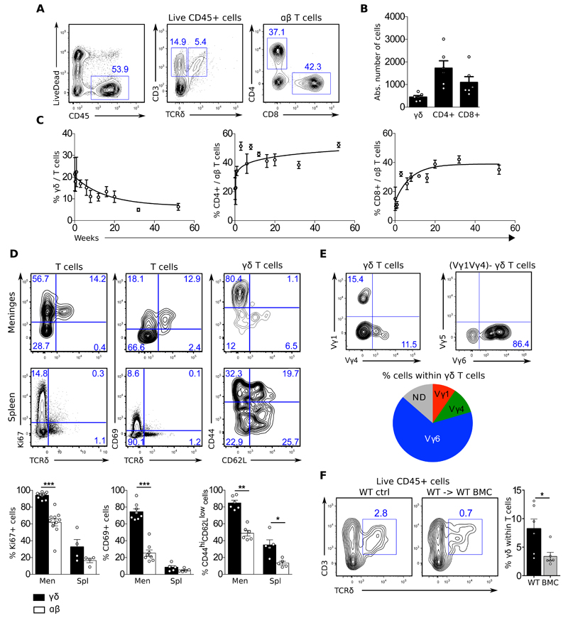
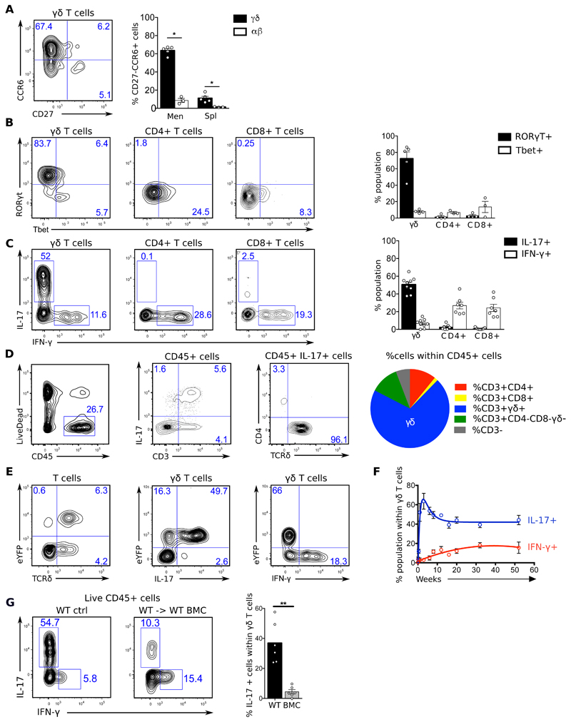
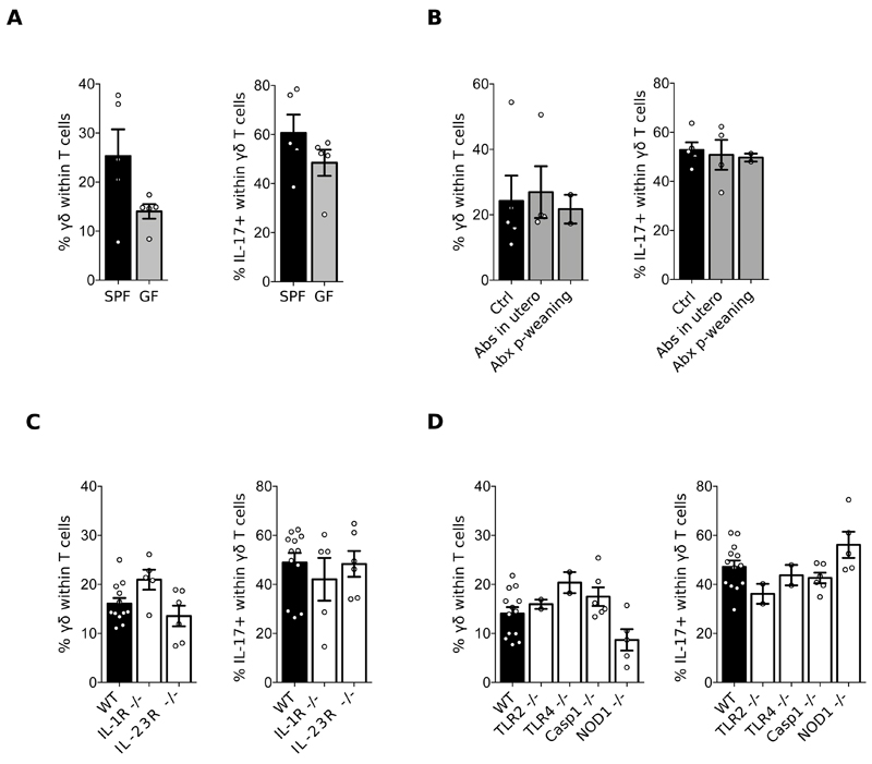
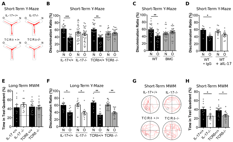
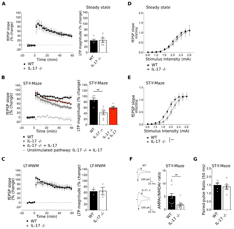
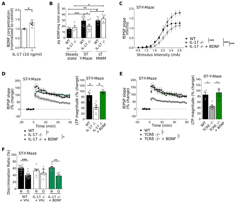
The notion of "immune privilege" of the brain has been revised to accommodate its infiltration, at steady state, by immune cells that participate in normal neurophysiology. However, the immune mechanisms that regulate learning and memory remain poorly understood. Here, we show that noninflammatory interleukin-17 (IL-17) derived from a previously unknown fetal-derived meningeal-resident γδ T cell subset promotes cognition. When tested in classical spatial learning paradigms, mice lacking γδ T cells or IL-17 displayed deficient short-term memory while retaining long-term memory. The plasticity of glutamatergic synapses was reduced in the absence of IL-17, resulting in impaired long-term potentiation in the hippocampus. Conversely, IL-17 enhanced glial cell production of brain-derived neurotropic factor, whose exogenous provision rescued the synaptic and behavioral phenotypes of IL-17-deficient animals. Together, our work provides previously unknown clues on the mechanisms that regulate short-term versus long-term memory and on the evolutionary and functional link between the immune and nervous systems.


Conflict of interest statement

Competing interests The authors declare that they have no conflict of interest.

Figures

Figure 1
Figure 1. Foetal derived γδ T cells infiltrate the meninges from birth.
Meningeal cell suspensions were prepared from 8-20 weeks-old C57BL/6J WT and Il17aCre R26ReYFP mice (A-B; D-E), 0-52 weeks-old C57BL/6J WT mice (C), and 20 weeks-old WT and WT → WT bone marrow chimeras (BMC) mice (G). Samples were analysed for the expression of indicated surface (CD45, CD3, TCRδ, CD4, CD8, Vγ1, Vγ4, Vγ5, Vγ6, CD27 and CCR6) and intracellular (RORgt, Tbet, IL-17 and IFN-γ) markers. Live cells were gated using LiveDead Fixable Viability Dye as shown in A. Dot plots represents cell populations from indicated gates. Histograms depicts percentages or absolute numbers from indicated populations. Meningeal spaces were pooled from 4 mice. Spleens were analysed from individual mice. Results are representative of 4-7 independent experiments. Error bars, mean + s.e.m. * P<0.05, **P<0.01, ***P<0.001 as calculated by student T-test (parametric) or Mann-Whitney test (non parametric).
Figure 2
Figure 2. Meningeal γδ T cells are biased towards IL-17 production.
Meningeal cell suspensions were prepared from 8-20 weeks-old C57BL/6J WT and Il17aCre R26ReYFP mice (A-E), 0-52 weeks-old C57BL/6J WT mice (G), and 20 weeks-old WT and WT → WT bone marrow chimeras (BMC) mice (F). Samples were analysed for the expression of indicated surface (CD45, CD3, TCRδ, CD4, CD8, Vγ1, Vγ4, Vγ5, Vγ6, CD27 and CCR6) and intracellular (RORgt, Tbet, IL-17 and IFN-γ) markers. Live cells were gated using LiveDead Fixable Viability Dye as shown in A. Dot plots represents cell populations from indicated gates. Histograms depicts percentages or absolute numbers from indicated populations. Meningeal spaces were pooled from 4 mice. Spleens were analysed from individual mice. Results are representative of 4-7 independent experiments. Error bars, mean + s.e.m. * P<0.05, **P<0.01, as calculated by Mann-Whitney test.
Figure 3
Figure 3. Meningeal γδ T cell homeostasis is independent of inflammatory signals
Cell suspensions were prepared from the meninges of 8-12 weeks-old C57BL/6J WT mice, bred in a Specific Pathogen Free (SPF) (A-D) versus Germ Free (GF) (A) environment, treated or not with an antibiotic cocktail (B), and compared to IL-1R-/-, IL-23R-/- (C), TLR2-/-, TLR4-/-, Caspase 1-/- and NOD1-/- mice (D). Percentages of γδ T cells and IL-17 producers were analysed by FACS as illustrated in Fig. 1. Results are representative of 2-4 independent experiments.
Figure 4
Figure 4. γδ T cells producing IL-17 are required for short-term memory.
(A) Representative track line from indicated animals exploring the short-term Y-maze. (B-D) Cognitive performance in the short-term Y-maze evaluated by discrimination ratio between the novel arm (N) versus the other arm (O) of IL-17-/-, TCRδ-/- compared to respective littermate controls (n=21-32) (B), WT → WT bone marrow chimeras (BMC) mice (n=23-27) (C) and WT after intra-cerebro-ventricular injection of isotype control (IgG) or anti-IL-17 (aIL-17) (n=10-12) (D). (E) Percentages of swimming time in the test quadrant of IL-17-/- and TCRδ-/- and respective littermate controls during the probe test of the long-term Morris Water Maze (MWM) (n=10-14). (F) Cognitive performance in the long-term Y-maze evaluated by discrimination ratio between the novel arm (N) versus the other arm (O) of IL-17-/-, TCRδ-/- compared to respective littermate controls (n=10-16). (G) Representative track line from indicated animals exploring the short-term Morris Water Maze (MWM) during the probe test, after a training phase with a platform in the lower left quadrant of the pool. (H) Corresponding percentages of time spent in the test quadrant of IL-17-/- and TCRδ-/- and respective littermate controls (n=8-16). Results are representative of 2-3 independent experiments in male mice. Error bars, mean + s.e.m. **P<0.05; **P<0.0; ***P<0.001. Paired Student’s t-test and One-way ANOVA followed by Bonferroni’s multiple comparison test were used to analyse discrimination ratio (%) and Time in quadrant (%) respectively.
Figure 5
Figure 5. IL-17 modulates synaptic plasticity and AMPA/NMDA ratio upon a short-term memory task.
(A-C) Time course (left panels) and magnitude (right panels) of LTP induced by theta-burst stimulation (TBS) in hippocampal slices from WT and IL-17-/- mice at steady state (A), after training in the short-term Y-Maze (B) and after training in the long-term MWM (C). When indicated, hippocampal slices from IL-17-/- mice were supplemented with IL-17 (10ng/mL) (n=3-7, Kruskal-Wallis test followed by Dunn's multiple comparisons test). (D-E) Input/Output (I/O) curves corresponding to the fEPSP slope evoked by different stimulation intensities (0.8-2.8 mA) of IL-17-/- compared to WT mice at steady state (D) and after training in the short-term Y-maze test (E) n=5-7, F-test. (F) Representative traces of EPSCs recorded at −70 mV and + 40 mV in neurons from WT and IL-17 -/- mice after training in the short-term Y-maze (left panels). Arrows indicate the amplitudes considered to calculate AMPAR/NMDAR ratio, depicted in the right panel. n=11-12, unpaired T-test. (G) Paired Pulse Facilitation, EPSCs at 50 ms interpulse intervals in WT and IL-17-/- after Y-maze (n=5-7, Mann-Whitney test). Data are mean ± s.e.m. **P<0.01, ***P<0.001.
Figure 6
Figure 6. IL-17 promotes glial Brain-Derived Neurotrophic Factor (BDNF) production.
(A) BDNF concentration in mixed glial cultures supplemented with IL-17 normalized to the control condition (n=4-6, Mann-Whitney test). (B) BDNF concentration in the hippocampus at steady state, after short-term Y-Maze test and after long-term MWM test (n=4-9, Mann-Whitney test). (C) I/O curves corresponding to the fEPSP slope evoked by different stimulation intensities (0.8-3.0 mA) of WT, IL-17-/- IL-17-/- supplemented with BDNF (30 ng/mL) after short-term Y-maze test (n=5-6, F-test). (D-E) Time course (left panel) and magnitude (right panel) of LTP induced by TBS in CA1 region of hippocampal slices of (D) WT, IL-17 -/- and IL-17-/- supplemented with BDNF (30 ng/mL) and (E) WT, TCRδ-/- and TCRδ-/- supplemented with BDNF (30 ng/mL) after short-term Y-Maze test (n=4-7, Kruskal-Wallis test followed by Dunn's multiple comparisons test.). Raw data from IL-17 -/- in panel C and D are adapted from Fig 3B and E. Data from WT is the same in panels D and E. (E) Cognitive performance in Y-Maze evaluated by discrimination ratio between N versus O of WT and IL-17-/- mice tested after ICV injection of PBS (vehicle, vhc) or BDNF (n=8-16, Paired Student's t-test). Results are representative of 2-5 independent experiments. Data are mean ± s.e.m, *P<0.05; **P<0.01; ***P<0.001.

Comment in

  • IL-17: an immune mnemonic.
    Bordon Y. Bordon Y. Nat Rev Immunol. 2019 Dec;19(12):720-721. doi: 10.1038/s41577-019-0237-1. Nat Rev Immunol. 2019. PMID: 31641283 No abstract available.

References

    1. Kipnis J, Gadani S, Derecki NC. Pro-cognitive properties of T cells. Nat Rev Immunol. 2012;12:663–669. - PMC - PubMed
    1. Weaver CT, Harrington LE, Mangan PR, Gavrieli M, Murphy KM. Th17: an effector CD4 T cell lineage with regulatory T cell ties. Immunity. 2006;24:677–688. - PubMed
    1. Ribot JC, deBarros A, Pang DJ, Neves JF, Peperzak V, Roberts SJ, Girardi M, Borst J, Hayday AC, Pennington DJ, Silva-Santos B. CD27 is a thymic determinant of the balance between interferon-gamma- and interleukin 17-producing gammadelta T cell subsets. Nat Immunol. 2009;10:427–436. - PMC - PubMed
    1. Jensen KD, Su X, Shin S, Li L, Youssef S, Yamasaki S, Steinman L, Saito T, Locksley RM, Davis MM, Baumgarth N, et al. Thymic selection determines gammadelta T cell effector fate: antigen-naive cells make interleukin-17 and antigen-experienced cells make interferon gamma. Immunity. 2008;29:90–100. - PMC - PubMed
    1. Munoz-Ruiz M, Ribot JC, Grosso AR, Goncalves-Sousa N, Pamplona A, Pennington DJ, Regueiro JR, Fernandez-Malave E, Silva-Santos B. TCR signal strength controls thymic differentiation of discrete proinflammatory gammadelta T cell subsets. Nat Immunol. 2016;17:721–727. - PMC - PubMed

Publication types