. 2019 Oct 11;9(1):14694.
doi: 10.1038/s41598-019-51022-1.

Brain lateralization probed by water diffusion at the atomic to micrometric scale

Affiliations

Brain lateralization probed by water diffusion at the atomic to micrometric scale

F Natali et al. Sci Rep. .

Abstract

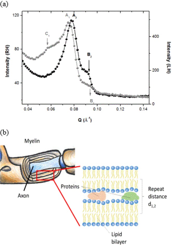
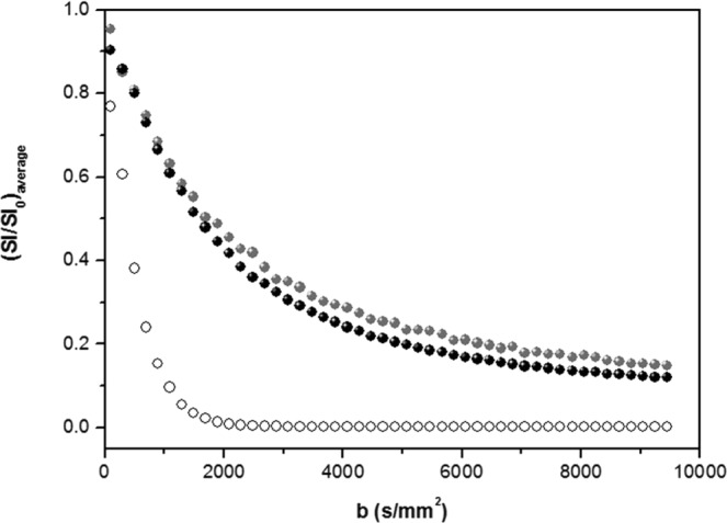
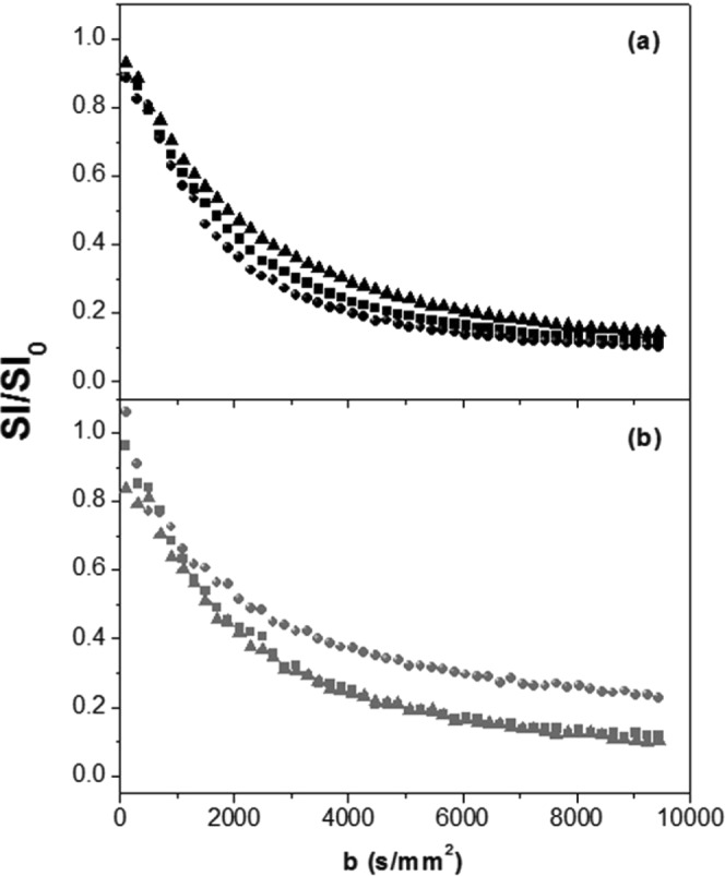
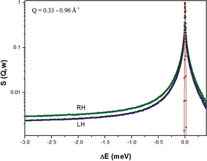
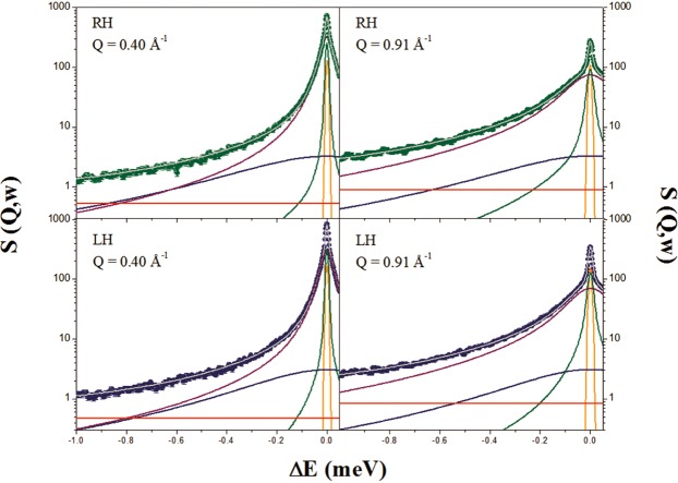
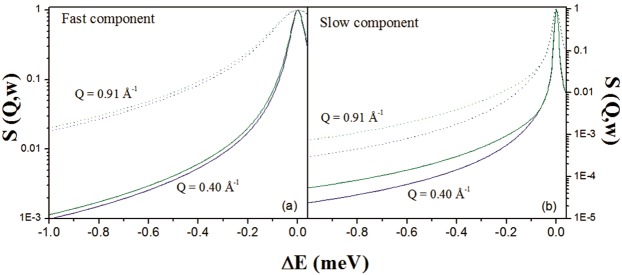
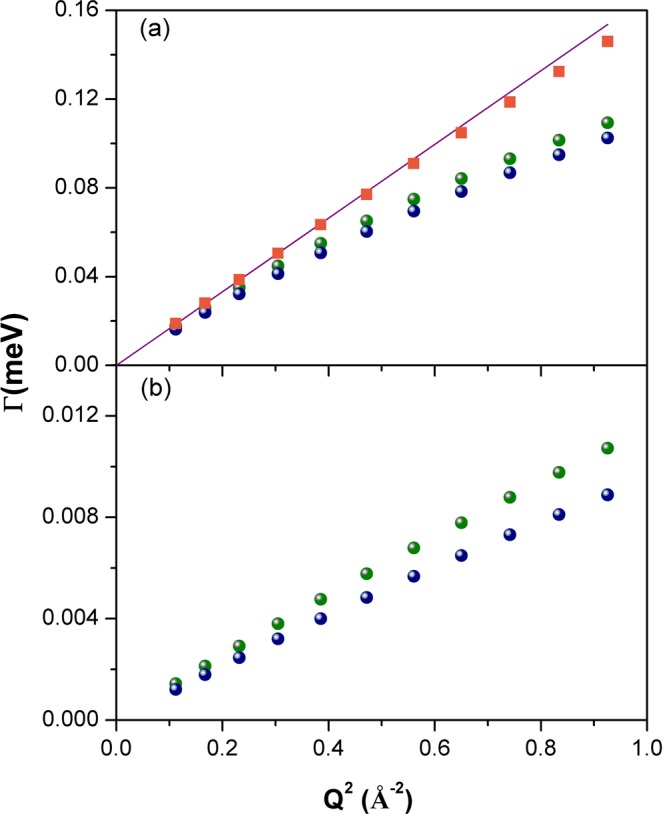
Combined neutron scattering and diffusion nuclear magnetic resonance experiments have been used to reveal significant interregional asymmetries (lateralization) in bovine brain hemispheres in terms of myelin arrangement and water dynamics at micron to atomic scales. Thicker myelin sheaths were found in the left hemisphere using neutron diffraction. 4.7 T dMRI and quasi-elastic neutron experiments highlighted significant differences in the properties of water dynamics in the two hemispheres. The results were interpreted in terms of hemisphere-dependent cellular composition (number of neurons, cell distribution, etc.) as well as specificity of neurological functions (such as preferential networking).


Conflict of interest statement

The authors declare no competing interests.

Figures

Figure 1
Figure 1
Panel a: Diffraction patterns obtained for the RH (black symbols) and LH (gray symbols) at 300 K measured on D16 at ILL. Second order Bragg peaks are found at A1 = 0.078 Å−1 (RH) and A2 = 0.074 Å−1 (LH). B1 and B2 represent less pronounced first (1st) or second (2nd) order Bragg peaks at Q = 0.093 Å−1 (RH) and Q = 0.088 Å−1 (LH). Additional reflection (C1) is observed in LH at Q = 0.057 Å−1. Panel b: Sketch of a myelin sheath and extraction of the multilamellar lipidic structure with attribution of the repeat distance d1 or d2.
Figure 2
Figure 2
dMRI signal decay of RH (black symbols) and LH (gray symbols) averaged over the three orthogonal directions; for comparison, the averaged signal decay of free water (deionized water Milli-Q, open symbols) is also shown.
Figure 3
Figure 3
dMRI signal decay of RH (panel a) and LH (panel b) normalized to signal decay at b = 0.5 s.mm−2, for three orthogonal directions (x: squares, y: circles and z: triangles).
Figure 4
Figure 4
Q-binned QENS intensities measured on IN5 of RH (green) and LH (blue) at 300 K. For completeness, the instrumental resolution function is also shown (red).
Figure 5
Figure 5
Example of QENS spectra of RH (green) and LH (blue) samples at Q = 0.40 Å−1 and Q = 0.91 Å−1 measured on IN5 at 300 K. The different scattering contributions, convoluted with instrumental resolution, are shown: experimental data (symbols), total fit (gray), elastic peak (orange), broad Lorentzian SCH2(Q, ω) (blue), fast water component (purple), slow water component (green) and background (red).
Figure 6
Figure 6
Contributions of the fast (panel a) and slow (panel b) water pool to the global QENS signal. RH: green; LH: blue; solid lines: Q = 0.40 Å−1; dotted lines: Q = 0.91 Å−1.
Figure 7
Figure 7
FWHM (Γ) of the translational motion for bulk-like water component (a) and restricted water component (b), for RH (green symbols), LH (blue symbols) and pure water (orange symbols), as a function of Q2, at 300 K. A Fick’s law function (purple line) has also been reported.

References

    1. Kandel, E. R., Schwartz, J. H. & Jessel, T. M. Principi di Neuroscienza. (ed. Casa Editrice Ambrosiana Milano) (1994).
    1. Pakkenberg B, Gundersen H. Neocortical neuron numbers in humans: effect of sex and age. J. Comp. Neurol. 1997;384:312–320. doi: 10.1002/(SICI)1096-9861(19970728)384:2<312::AID-CNE10>3.0.CO;2-K. - DOI - PubMed
    1. Pakkenberg B, et al. Aging and the human neocortex. Exper. Geront. 2003;38:95–99. doi: 10.1016/S0531-5565(02)00151-1. - DOI - PubMed
    1. Williams RW, Herrup K. The control of neuron number. Ann Rev Neuroscience. 1988;11:423–453. doi: 10.1146/annurev.ne.11.030188.002231. - DOI - PubMed
    1. Ross ED, Monnot M. Neurology of affective prosody and its functional-anatomic organization in right hemisphere. Brain Lang. 2008;104(1):51–74. doi: 10.1016/j.bandl.2007.04.007. - DOI - PubMed

Publication types