. 2019 Oct 17;179(3):644-658.e13.
doi: 10.1016/j.cell.2019.09.028. Epub 2019 Oct 10.

Segmented Filamentous Bacteria Prevent and Cure Rotavirus Infection

Affiliations

Segmented Filamentous Bacteria Prevent and Cure Rotavirus Infection

Zhenda Shi et al. Cell. .

Abstract

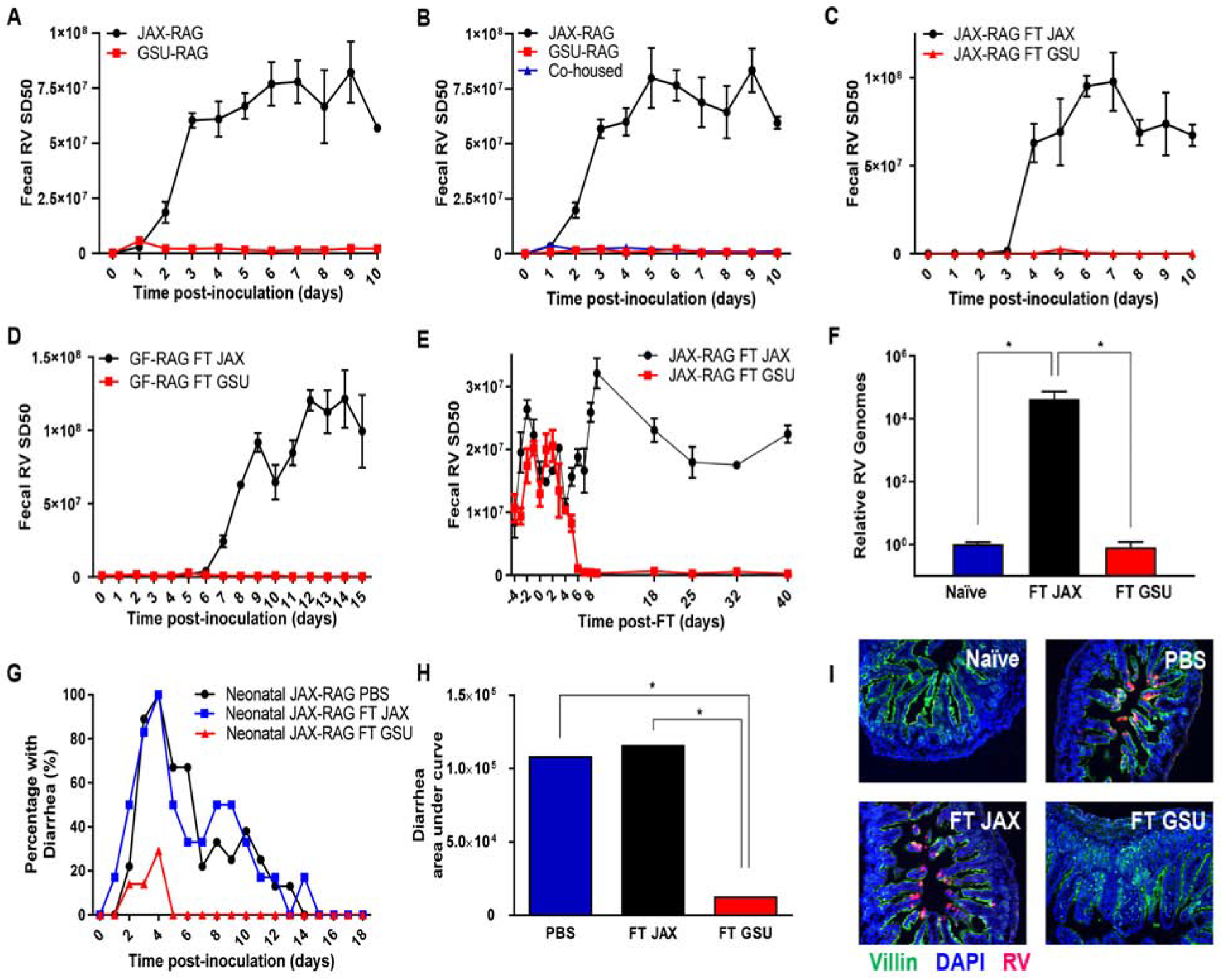
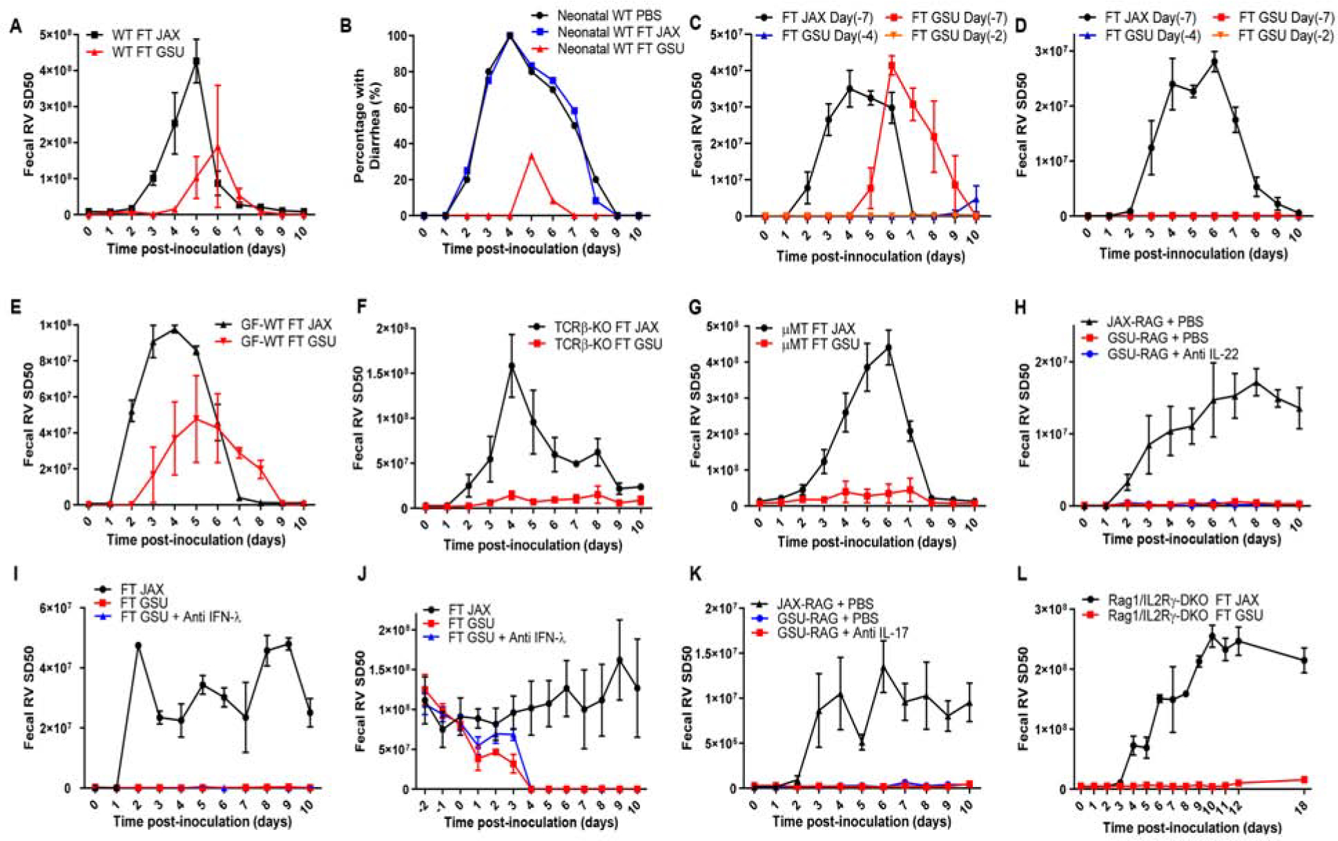
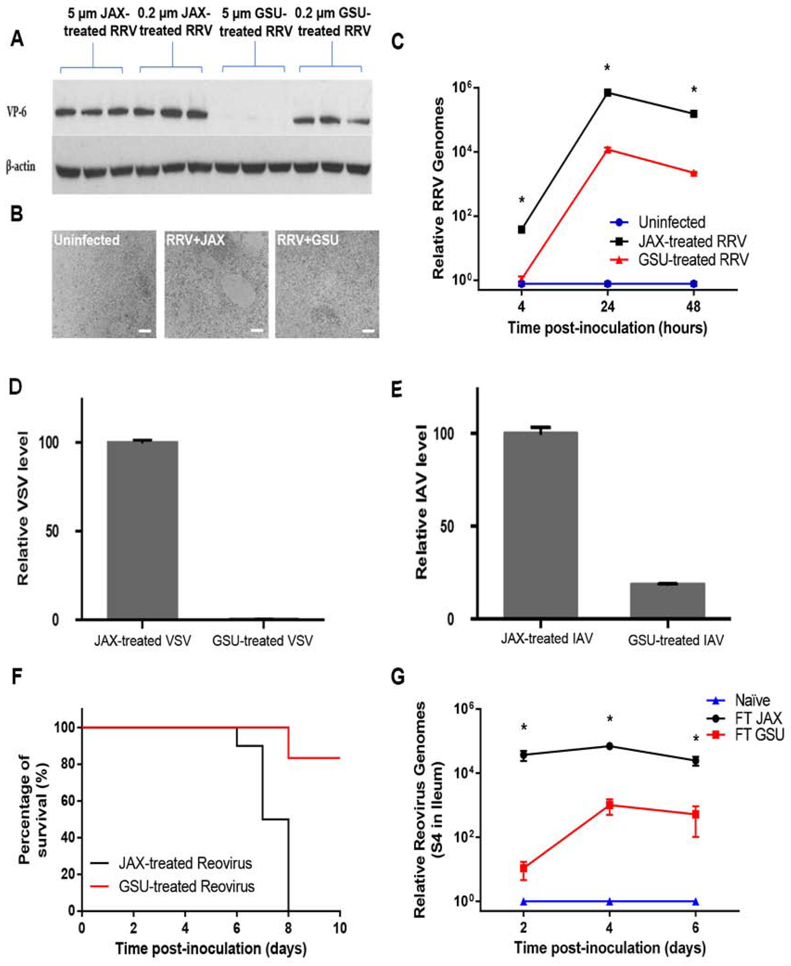
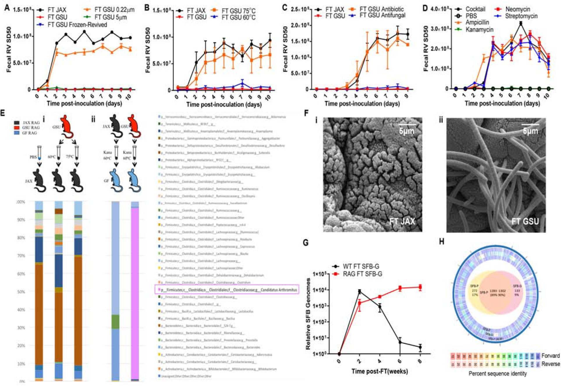
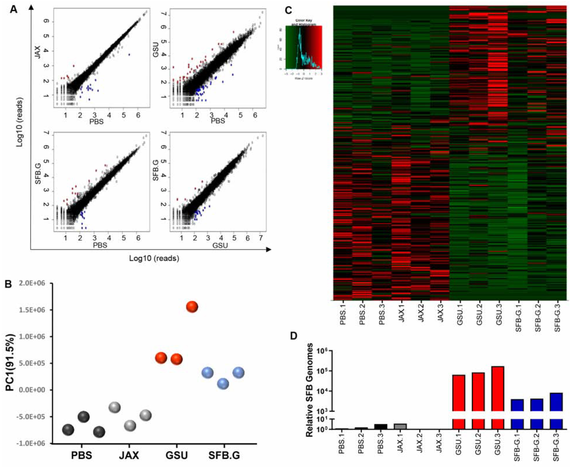
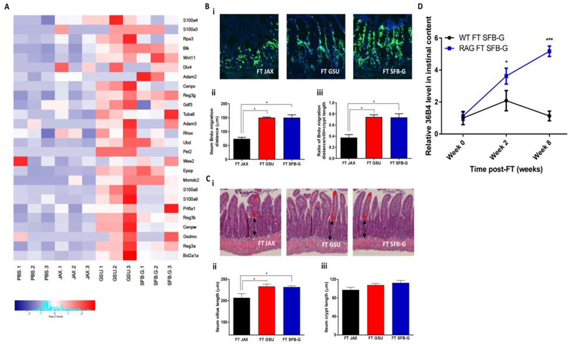
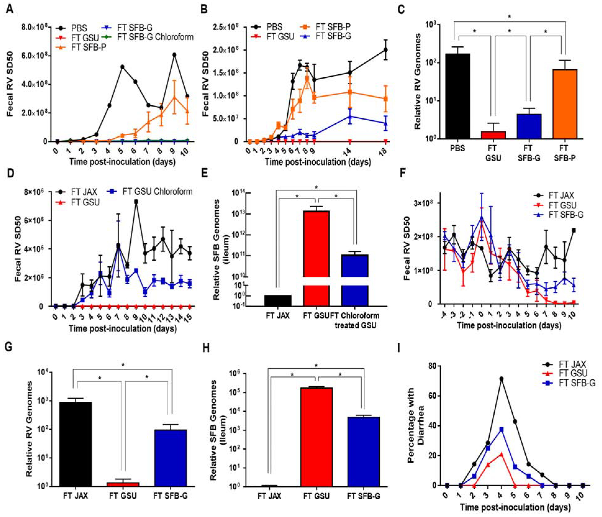
Rotavirus (RV) encounters intestinal epithelial cells amidst diverse microbiota, opening possibilities of microbes influencing RV infection. Although RV clearance typically requires adaptive immunity, we unintentionally generated RV-resistant immunodeficient mice, which, we hypothesized, reflected select microbes protecting against RV. Accordingly, such RV resistance was transferred by co-housing and fecal transplant. RV-protecting microbiota were interrogated by heat, filtration, and antimicrobial agents, followed by limiting dilution transplant to germ-free mice and microbiome analysis. This approach revealed that segmented filamentous bacteria (SFB) were sufficient to protect mice against RV infection and associated diarrhea. Such protection was independent of previously defined RV-impeding factors, including interferon, IL-17, and IL-22. Colonization of the ileum by SFB induced changes in host gene expression and accelerated epithelial cell turnover. Incubation of RV with SFB-containing feces reduced infectivity in vitro, suggesting direct neutralization of RV. Thus, independent of immune cells, SFB confer protection against certain enteric viral infections and associated diarrheal disease.

Keywords: fecal transplant; germ-free mice; infectious diarrhea; microbiota-virus interactions; rotavirus; segmented filamentous bacteria; viral gastroenteritis.


Conflict of interest statement

Declaration of Interests

The authors declare no competing interests.

Figures

Figure 1.
Figure 1.. Transmissible Microorganisms Protect Immune-Deficient Mice Against Rotavirus Infection
Rag1-KO mice were orally inoculated with RV, EC strain. Feces were collected daily, and fecal RV antigen quantified by ELISA. (A) Comparison of mice from two colonies of Rag1-KO mice (GSU and JAX). (B) JAX, GSU, and JAX co-housed with GSU, Rag1-KO mice. (C) JAX Rag1-KO mice received fecal transplants (FT) from JAX or GSU Rag1-KO mice one week prior to RV inoculation. (D) Germ-free (GF) Rag1-KO mice received FT from JAX or GSU Rag1-KO mice one week prior to RV inoculation. (E–F) JAX Rag1-KO with established chronic RV infections received FT from JAX or GSU Rag1-KO mice. Panel E shows fecal RV antigens, while panel F shows levels of intestinal RV genomes quantified by qRT-PCR at day 62 post-transplantation. (G–I) Seven-day-old Rag1-KO mice pups received PBS or FT from JAX or GSU Rag1-KO mice 6 d and 1 d prior to RV inoculation. Panels G and H show the incidence of diarrhea, while I shows RV antigen by fluorescence microscopy 5 weeks post-inoculation. Data shown are means ± SEM of individual experiments and were significant by two-way analysis of variance (ANOVA); A; n = 3, P < 0.0001. B; n = 3–4, P < 0.0001. C-D; n = 4, P < 0.0001. E-F; n = 6, P < 0.0001. G, FT GSU differed significantly from both FT JAX and the control (FT WT) groups by one-way ANOVA, n = 12, *, P < 0.0001; H, FT GSU differed significantly from both FT JAX and the control (PBS) groups by χ2 test, n=12, *, P < 0.01. I, representative images (n= 4 mice per condition). Each experiment was performed 2–3 times and yielded an identical pattern of results.
Figure 2.
Figure 2.. Bacterial Constituents of GSU RAG Feces Partially Protect Immune-Competent Mice from RV
Mice were orally inoculated with RV, EC strain. Feces were collected daily, and fecal RV antigen was quantified by ELISA. (A) WT C57BL/6J mice received FT from JAX or GSU Rag1-KO mice one week prior to RV inoculation. (B) Seven-day-old WT C57BL/6J mice pups received PBS or FT from JAX or GSU Rag1-KO mice twice 6 d and 1 d prior to RV inoculation. Panels B shows the incidence of diarrhea. (C–D) (C) Eight-week-old or (D) Three-week-old WT mice received FT from JAX or GSU Rag1-KO mice. Mice were then inoculated 2, 4, or 7 days later with RV (7-days for recipients of JAX FT). (E) GF WT C57BL/6J mice received FT from JAX or GSU Rag1-KO mice one week prior to RV inoculation. (F) TCRβ-KO mice received FT from JAX or GSU Rag1-KO mice one week prior to RV inoculation. (G) μMT (Ighm-KO) mice received FT from JAX or GSU Rag1-KO mice one week prior to RV inoculation. (H) JAX or GSU colony of Rag1-KO mice were inoculated intraperitoneally with PBS or IL-22 neutralizing antibody (150 μg daily for 2 days) neutralizing antibody shortly before RV inoculation. (I) JAX colony of Rag1-KO mice were inoculated intraperitoneally daily with PBS or IFN-λ neutralizing antibody (1 μg per gram of body weight/day) from day −1 to day 10 following RV inoculation. (J) RV chronically infected JAX Rag1-KO mice received FT with GSU mice microbiota or GSU microbiota with intraperitoneal inoculation of IFN-λ neutralizing antibody (1μg per gram of body weight/day) from day −1 to day 10 post-FT. (K) JAX or GSU colony of Rag1-KO mice were inoculated intraperitoneally with PBS or IL-17 neutralizing antibody (100μg daily for 2days) neutralizing antibody shortly before RV inoculation. (L) ILC-deficient (Rag1/IL2Rγ-DKO) received FT from JAX or GSU Rag1-KO mice one week prior to RV inoculation. Data shown are means ± SEM (two-way ANOVA). A; n = 3, P < 0.01. C; n=3, P < 0.0001. D; n=5, P < 0.0001. E; n = 4, P < 0.05. F; n = 3, P < 0.0001. G; n = 3, P < 0.0001. H-I; n = 4, P < 0.0001. J; n=3, P < 0.0001. K; n=3, P < 0.01. L; n = 4, P < 0.0001. B, FT GSU differed significantly from both FT JAX and the control (FT WT) groups by one-way ANOVA, n = 12, *, P < 0.01. Each experiment was performed 2–3 times and yielded an identical pattern of results.
Figure 3.
Figure 3.. GSU-RAG Microbiota May Directly Interact with Viruses to Reduce Infectivity
The viruses tested were incubated with 5 μm filtered JAX or GSU Rag1-KO mice fecal samples at 37°C. The mixtures were then filtered through 0.2 μm filters to remove the bacteria cells and used for in vitro challenges. (A-C) RRV was pre-incubated with 5 μm or 0.2 μm filtered JAX or GSU Rag1-KO mice fecal samples at 37°C for 4 hours prior to inoculating HT-29 cells in vitro. (A) Immunoblotting for RV VP6 at 20 hours after infection with RRV. (B) Microscopic analysis of HT-29 cells at 20 hours after infection with treated RRV (5 μm filtered JAX or GSU Rag1-KO mice fecal samples), scale bar: 100 μm. (C) qRT-PCR results at the time points shown after infection with treated RRV (5 μm filtered JAX or GSU Rag1-KO mice fecal samples). (D-E) Effect of fecal microbiota (5 μm filtered JAX or GSU Rag1-KO mice fecal samples) on VSV (D) and IAV WSN (E) infection of BEAS-B cells 24 hours post-infection in vitro. (F) Survival of IFN-αR-KO mice following inoculation with reovirus treated with either 5 μm-filtered JAX or GSU Rag1-KO mice fecal samples (37°C, 4 hours). (G) qRT-PCR assay of reovirus RNA in the small intestine of Rag1-KO mice following transplant of JAX or GSU Rag1-KO mice fecal samples 1 week prior to oral inoculation. Results are shown as mean ± SEM (two-way ANOVA). A-B, representative images (n= 3 per condition). C; GSU treated RRV differed significantly from control (JAX treated RRV) group by two-way ANOVA, n = 4, *, P < 0.0001. D; n = 4, P < 0.0001. E; n = 4, P < 0.0001. F; χ2 test, n = 10, P < 0.0001. G; FT GSU group differed significantly from control (FT JAX) group by two-way ANOVA, n = 3, *, P < 0.0001. Each experiment was performed 2–3 times and yielded an identical pattern of results.
Figure 4.
Figure 4.. RV-Resistance Correlates with Transfer of Segmented Filamentous Bacteria (SFB)
JAX Rag1-KO mice were orally inoculated with RV, EC strain. Feces were collected daily, and fecal RV antigen was quantified by ELISA. (A) JAX Rag1-KO mice received FT from JAX or GSU Rag1-KO mice with fresh, −80°C revived, 0.22 μm filtered, or 5 μm filtered fecal samples for 1 week prior to RV inoculation. (B) JAX Rag1-KO mice received FT from JAX or GSU Rag1-KO mice with untreated or heat-treated (60°C or 75°C for 10 minutes) fecal samples for 1 week prior to RV inoculation. (C) JAX Rag1-KO mice received FT with JAX or GSU Rag1-KO mice fecal samples that were treated with an antibiotic cocktail or anti-fungal cocktail (37°C for 4 hours) 1 week prior to RV inoculation. (D) JAX Rag1-KO mice received FT with PBS, ampicillin, kanamycin, neomycin, streptomycin, or all four antibiotics combined (37°C for 4 hours) GSU Rag1-KO mice fecal samples 1 week prior to RV inoculation. (E) (i) 16s rRNA sequencing results of the conventional JAX Rag1-KO mice that received PBS or 60°C-, or 75°C-treated GSU Rag1-KO mice fecal samples. (ii) Fecal 16s rRNA sequencing results of GF Rag1-KO mice that were transplanted with diluted kanamycin-treated, heat-treated (60°C for 10 minutes) JAX or GSU Rag1-KO mice fecal samples. (F) Scanning electron micrographs of ileal sections from mice in E-ii. Those receiving GSU Rag1-KO were abundantly colonized by SFB. (G) Fecal SFB levels by qPCR in WT or Rag-1KO mice following FT with GSU-SFB. (H) Genomes of SFB strain in E-ii (SFB-G) and reference strain provided by Pasteur Institute (SFB-P) were sequenced and compared to SFB-P genome sequence data in the NCBI. Outer ring: Pan-genome analysis of forward and reverse assembly summary. Inner ring: Venn diagram representing the number of sequences unique or shared between the two assembled strains (90%–95% identity between the two strains at the protein level). Results are shown as mean ± SEM (two-way ANOVA). A-B; n = 4, P < 0.0001. C-D; n = 3, P < 0.0001. H; n=4, P < 0.01. Experiments A-D were performed 2–3 times and yielded an identical pattern of results.
Figure 5.
Figure 5.. GSU-RAG Microbiota and SFB-G Alter Host Gene Expression
Rag1-KO mice were orally administered PBS or feces from JAX-RAG, GSU-RAG, or SFB-G-monoassociated mice. Ileum was harvested at 2 weeks post-FT for RNAseq analysis. (A) Linear gene expression comparison. (B) PCA plot of PC1. (C) Heatmap of genes significantly altered (P < 0.05) by two-fold in any group relative to the PBS control group. (D) Levels of SFB genomes in the same ileal samples determined by real-time PCR. N=3 mice per condition.
Figure 6.
Figure 6.. GSU-RAG Microbiota and SFB-G Increase Gut Epithelial Proliferation and Shedding
Rag1-KO mice or WT mice were orally transplanted with feces from JAX-RAG, GSU-RAG, or SFBG-monoassociated mice. Ileum was harvested at 1 week. (A) Heatmap of genes, which are related with cell cycle, cell phase, cell proliferation, and cell death that are significantly altered (P < 0.05) by two-fold in any group relative to the PBS control group. N=3 mice per condition. (B) BrdU was inoculated intraperitoneally into transplanted Rag1-KO mice 24 hours before harvesting the tissues. (C) (i) H&E staining of Rag1-KO mice ileum. (ii) Villus length, indicated by the red arrow, was quantified. (iii) Crypt length, indicated by the black arrow, was quantified. (D) Ileum contents were harvested at 0, 2, and 4 weeks post-transplantation, and cells were quantified by qPCR. Results are shown as mean ± SEM (Student’s t test). A; n = 6, *, P < 0.001. B; n = 6, *, P < 0.05. C; n = 4, *, P < 0.05. D; n = 4, *, P < 0.05; ***, P < 0.001. Each experiment was performed two times and yielded an identical pattern of results.
Figure 7.
Figure 7.. SFB Confers Strain-Specific Resistance to RV Infection
Rag1-KO mice were orally inoculated with RV, EC strain. Feces were collected daily, and fecal RV antigen was quantified by ELISA. Ileum or fecal samples were collected for assaying SFB or RV levels by qPCR. (A) GF Rag1-KO mice received FT from PBS, GSU Rag1-KO mice, Pasteur-SFB (SFB-P), SFB isolated from GSU Rag1-KO mice (SFB-G), or chloroform-treated (3% at RT for 1 hour) SFB-G for 1 week prior to RV inoculation. (B-C) (B) Conventional JAX Rag1-KO mice received FT from PBS, GSU Rag1-KO mice, SFB-P, or SFB-G for 1 week prior to RV inoculation. (C) Relative RV genome levels in the recipient mice. (D-E) (D) Conventional JAX Rag1-KO mice received FT from PBS, GSU, or chloroform treated (3% at RT for 1 hour) GSU Rag1-KO mice for 1 week prior to RV inoculation. (E) Ileum SFB level by q-PCR. (F-H) SFB-G significantly reduced RV shedding in chronically infected recipients. (F) Rag1-KO mice chronically infected with RV received FT from JAX, GSU Rag1-KO mice, or SFB-G. (G) qRT-PCR results of ileum RV genome levels at day 10 post-transfer. (H) Ileum SFB levels 1-week post FT. (I) Seven-day-old WT C57BL/6 mice pups received FT from JAX, GSU, or SFB-G mono-associated Rag1-KO mice 6 d and 1 d prior to RV inoculation. Results are shown as mean ± SEM (two-way ANOVA). A; n = 3, P < 0.0001. B; n = 3, P < 0.0001. C; Student’s t test, n = 3, *, P < 0.01. D; n = 3, P < 0.01. E; Student’s t test, n = 3, *, P < 0.05. F; n = 3, P < 0.001. G-H; Student’s t test, n = 3, *, P < 0.01. I; Both FT GSU and FT SFB-G differed significantly from control (FT JAX) group by one-way ANOVA, n = 8 mice, P < 0.05. Each experiment was performed 2–3 times and yielded an identical pattern of results.

Comment in

References

    1. Aguilera ER, Nguyen Y, Sasaki J, and Pfeiffer JK (2019). Bacterial Stabilization of a Panel of Picornaviruses. mSphere 4. - PMC - PubMed
    1. Baldridge MT, Lee S, Brown JJ, McAllister N, Urbanek K, Dermody TS, Nice TJ, and Virgin HW (2017). Expression of Ifnlr1 on Intestinal Epithelial Cells Is Critical to the Antiviral Effects of Interferon Lambda against Norovirus and Reovirus. J Virol 91. - PMC - PubMed
    1. Bankevich A, Nurk S, Antipov D, Gurevich AA, Dvorkin M, Kulikov AS, Lesin VM, Nikolenko SI, Pham S, Prjibelski AD, et al. (2012). SPAdes: a new genome assembly algorithm and its applications to single-cell sequencing. J Comput Biol 19, 455–477. - PMC - PubMed
    1. Barro M, and Patton JT (2007). Rotavirus NSP1 inhibits expression of type I interferon by antagonizing the function of interferon regulatory factors IRF3, IRF5, and IRF7. J Virol 81, 4473–4481. - PMC - PubMed
    1. Basu R, O’Quinn DB, Silberger DJ, Schoeb TR, Fouser L, Ouyang W, Hatton RD, and Weaver CT (2012). Th22 cells are an important source of IL-22 for host protection against enteropathogenic bacteria. Immunity 37, 1061–1075. - PMC - PubMed

Publication types

MeSH terms