Review
. 2020 Jun;13(6):1368-1383.
doi: 10.1016/j.jcmg.2019.07.015. Epub 2019 Oct 11.

How to Image Cardiac Amyloidosis: A Practical Approach

Affiliations
Review

How to Image Cardiac Amyloidosis: A Practical Approach

Sharmila Dorbala et al. JACC Cardiovasc Imaging. 2020 Jun.

Abstract

Cardiac amyloidosis (CA) is one of the most rapidly progressive forms of heart disease, with a median survival from diagnosis, if untreated, ranging from <6 months for light chain amyloidosis to 3 to 5 years for transthyretin amyloidosis. Early diagnosis and accurate typing of CA are necessary for optimal management of these patients. Emerging novel disease modifying therapies increase the urgency to diagnose CA at an early stage and identify patients who may benefit from these life-saving therapies. The goal of this review is to provide a practical approach to echocardiography, cardiac magnetic resonance, and radionuclide imaging in patients with known or suspected CA.

Keywords: CMR; PET; Tc-99m−PYP; amyloid tracers; cardiac amyloidosis; cardiac magnetic resonance; echocardiography; imaging; multimodality; pyrophosphate; radionuclide imaging.


Figures

FIGURE 1
FIGURE 1. Echocardiography in CA
In the context of a thick Left ventricle (LV), the following features should be considered red flags for cardiac amyloidosis (CA): (A) pericardial and/or pleural effusions, thick right ventricle, thick valves, thick interatrial septum, small LV cavity size with low stroke volume, and paradoxical low flow, low gradient aortic stenosis. (B) Advanced CA results in restrictive LV filling patterns with rapid E-wave deceleration time, a predominant S wave on pulmonary venous Doppler, and low myocardial relaxation velocities on tissue Doppler imaging (TDI) at the mitral annulus. Finally, amyloid infiltration impairs (C) global Longitudinal strain (GLS) characteristically with apical sparing of the LV apex, in contrast to a normal pattern, and severely reduced contractile function of the atrial myocardium, in contrast to a normal pattern. AS = aortic stenosis.
Figure 2
Figure 2. Cardiac Structural Changes Typically Indicate Advanced CA
Images of a 54-year-old African American man with heart failure who eventually succumbed to sepsis. (A) Echocardiogram, (B) technetium-99m (Tc-99m) -pyrophosphate (PYP) single-photon emission computed tomography/computed tomography (SPECT/CT) (strongly positive, grade 3 uptake), and (C) autopsy images are shown. A plasma cell dyscrasia was excluded, and genetic testing revealed a TTR gene mutation (Asp18Asn). Autopsy confirmed substantial transmural and diffuse amyloid deposition. (D) Low- and high-power sulfated Alcian Blue stain with blue/green staining amyloid and yellow/brown staining myocytes. Autopsy and histology images courtesy of Dr. Robert E. Padera. Abbreviation as in Figure 1.
FIGURE 3
FIGURE 3. A High Clinical Suspicion of ATTR CA Is Needed in Older Persons With Hypertension and Increased LV wall Thickness
Images of a 76-year-old man with a history of hypertension and progressive increase in LV wall thickness from (A) 10 mm in 2011 to (B) 18 mm in 2018. His cardiologist questioned whether this could be explained solely on the basis of hypertension. Echocardiography also revealed right ventricular wall thickening, raising the possibility of transthyretin (ATTR) CA. Serum and urine immunofixation studies and serum light chain levels were normal. (C and D) A Tc-99-PYP scan revealed grade 3 myocardial uptake, confirming ATTR CA. Abbreviations as in Figures 1 and 2.
FIGURE 4
FIGURE 4. CMR in CA
Cardiovascular magnetic resonance (CMR) provides characteristic imaging of the (A) structural changes and (B) powerful tissue characterization with features of high native T1, expanded extracellular volume (ECV), and late gadolinium enhancement (LGE) (diffuse, subendocardial, or transmural). (C) Post-gadolinium myocardial signal intensity changes characteristically with myocardial signal nulling before the blood pool signal in amyloidosis and vice versa in non-amyloid hearts. Abbreviation as in Figure 1.
FIGURE 5
FIGURE 5. Tc-99m–PYP in CA
Tc-99m –PYP planar and SPECT scans graded visually (grade 0, negative, to grade 3, strongly positive) and semi-quantitatively using the heart to contralateral lung uptake (HCL) ratio (normal, equivocal and diagnostic of ATTR). Abbreviations as in Figures 1 to 3.
FIGURE 6
FIGURE 6. A Negative Tc-99m–PYP Does Not Exclude CA
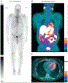
A 56-year-old African American man with a V122I TTR mutation presented with heart failure and an echocardiogram suspicious for CA. (A) A Tc-99m–PYP scan revealed grade 1 uptake (myocardial uptake less than rib uptake). Serum and urine immunofixation and serum light chain assay suggested light chain (AL) amyloidosis. An endomyocardial biopsy with mass spectroscopy confirmed Lambda AL CA. (B) Cardiac and partial whole body F-18-florbetapir positron emission tomography (PET/CT) images, as part of a research protocol, demonstrated intense myocardial and parotid uptake of F-18-florbetapir. He received a regimen of bortezomib, cyclophosphamide, and dexamethasone with complete hematological response. Abbreviations as in Figures 1 and 2.
Figure 7
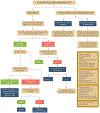
Figure 7. An Algorithmic Approach to Imaging-Based Evaluation of CA
CA may be considered in patients with heart failure as well as in patients without heart failure but with a predisposition to CA. MGUS = monoclonal gammopathy of uncertain significance; Pts = patients; other abbreviation as in Figure 1.
CENTRAL ILLUSTRATION
CENTRAL ILLUSTRATION. Imaging Targets in Cardiac Amyloidosis
Interstitial amyloid deposits (purple) thicken the myocardium and expand ECV; but, these features or voltage on electrocardiography cannot definitively distinguish infiltration from true myocardial hypertrophy. Targeted amyloid imaging with radiotracers are specific to image myocardial amyloid deposits. CMR = cardiac magnetic resonance; DPD = -3,3-diphosphono-1,2 propanodicarboxylic acid; ECV=extracellular volume; HMDP = hydroxymethylene diphosphonate; PiB = Pittsburgh B compound; PYP = pyrophosphate; SAP = serum amyloid P component; 99mTc = technetium-99m.

Comment in

References

    1. MerLini G, PaLLadini G. Light chain amyloidosis: the heart of the problem. HaematoLogica 2013;98:1492–5. - PMC - PubMed
    1. Ruberg FL, Berk JL. Transthyretin (TTR) cardiac amyloidosis. Circulation 2012;126:1286–300. - PMC - PubMed
    1. Glenner GG, Terry W, Harada M, Isersky C, Page D. Amyloid fibril proteins: proof of homology with immunoglobulin Light chains by sequence analyses. Science 1971;172:1150–1. - PubMed
    1. Gonzalez-Lopez E, Gallego-Delgado M, Guzzo-Merello G, et al. Wild-type transthyretin amyloidosis as a cause of heart failure with preserved ejection fraction. Eur Heart J 2015;36:2585–94. - PubMed
    1. Castano A, Narotsky DL, Hamid N, et al. Unveiling transthyretin cardiac amyloidosis and its predictors among elderly patients with severe aortic stenosis undergoing transcatheter aortic valve replacement. Eur Heart J 2017;38:2879–87. - PMC - PubMed

Publication types

MeSH terms

Supplementary concepts