. 2019 Sep 24:10:2244.
doi: 10.3389/fmicb.2019.02244. eCollection 2019.

Microbial Community Shifts Associated With the Ongoing Stony Coral Tissue Loss Disease Outbreak on the Florida Reef Tract

Affiliations

Microbial Community Shifts Associated With the Ongoing Stony Coral Tissue Loss Disease Outbreak on the Florida Reef Tract

Julie L Meyer et al. Front Microbiol. .

Abstract

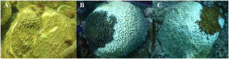
As many as 22 of the 45 coral species on the Florida Reef Tract are currently affected by stony coral tissue loss disease (SCTLD). The ongoing disease outbreak was first observed in 2014 in Southeast Florida near Miami and as of early 2019 has been documented from the northernmost reaches of the reef tract in Martin County down to Key West. We examined the microbiota associated with disease lesions and apparently healthy tissue on diseased colonies of Montastraea cavernosa, Orbicella faveolata, Diploria labyrinthiformis, and Dichocoenia stokesii. Analysis of differentially abundant taxa between disease lesions and apparently healthy tissue identified five unique amplicon sequence variants enriched in the diseased tissue in three of the coral species (all except O. faveolata), namely an unclassified genus of Flavobacteriales and sequences identified as Fusibacter (Clostridiales), Planktotalea (Rhodobacterales), Algicola (Alteromonadales), and Vibrio (Vibrionales). In addition, several groups of likely opportunistic or saprophytic colonizers such as Epsilonbacteraeota, Patescibacteria, Clostridiales, Bacteroidetes, and Rhodobacterales were also enriched in SCTLD disease lesions. This work represents the first microbiological characterization of SCTLD, as an initial step toward identifying the potential pathogen(s) responsible for SCTLD.

Keywords: Caribbean; coral microbiome; dysbiosis; scleractinian coral; white syndrome.


Figures

FIGURE 1
FIGURE 1
Stony coral tissue loss disease appearance in three coral species from the Florida Reef Tract: Montastraea cavernosa (A), Diploria labyrinthiformis (B), and Dichocoenia stokesii (C).
FIGURE 2
FIGURE 2
Principal component analysis of microbial community structure in disease lesions (DD), apparently healthy tissue on diseased corals (DH), and apparently healthy neighboring corals with no signs of disease (H).
FIGURE 3
FIGURE 3
Relative abundance of amplicon sequence variants, colored by Class, in undiseased neighboring corals, apparently healthy tissue far from the disease lesion, apparently healthy tissue near the disease lesion, and disease lesions in Montastraea cavernosa (A), Orbicella faveolata (B), Diploria labyrinthiformis (C), and Dichocoenia stokesii (D) with stony coral tissue loss disease.
FIGURE 4
FIGURE 4
The dispersion of beta diversity shown as the distance to the centroid in microbial communities from disease lesions (DD), apparently healthy tissue on diseased corals (DH), and apparently healthy neighboring corals with no signs of disease (H) in Montastraea cavernosa, Diploria labyrinthiformis, and Dichocoenia stokesii.
FIGURE 5
FIGURE 5
Relative abundance (RA) of the five amplicon sequence variants [Cyromorphaceae (A), Fusibacter (B), Planktotalea (C), Algicola (D), and Vibrio (E)] that were differentially abundant in disease lesions versus apparently healthy (non-lesion) tissues of Montastraea cavernosa (Mcav), Orbicella faveolata (Ofav), Diploria labyrinthiformis (Dlab), and Dichocoenia stokesii (Dsto). For display on a logarithmic scale, 0.0001 was added to all values to avoid taking the log of zero.
FIGURE 6
FIGURE 6
Relative abundance of 21 microbial families that were differentially abundant across coral tissue types: disease lesions, apparently healthy tissue near the lesions, apparently healthy tissue far from the lesions on diseased colonies, and neighboring undiseased colonies. Points represent the average relative abundance and error bars depict the standard error from analysis of all 62 coral samples. For display on a logarithmic scale, 0.001 was added to all values to avoid taking the log of zero.

References

    1. Apprill A., McNally S., Parsons R., Weber L. (2015). Minor revision to V4 region SSU rRNA 806R gene primer greatly increases detection of SAR11 bacterioplankton. Aquat. Microb. Ecol. 75 129–137. 10.3354/ame01753 - DOI
    1. Aronson R. B., Precht W. F. (2001). “White-band disease and the changing face of caribbean coral reefs,” in The Ecology and Etiology of Newly Emerging Marine Diseases, ed. Porter J. W. (Dordrecht: Springer Netherlands; ), 25–38. 10.1007/978-94-017-3284-0_2 - DOI
    1. Arotsker L., Kramarsky-Winter E., Ben-Dov E., Siboni N., Kushmaro A. (2015). Changes in the bacterial community associated with black band disease in a Red Sea coral, Favia sp., in relation to disease phases. Dis. Aquat. Organ. 116 47–58. 10.3354/dao02911 - DOI - PubMed
    1. Bayer T., Neave M. J., Alsheikh-Hussain A., Aranda M., Yum L. K., Mincer T., et al. (2013). The microbiome of the red sea coral Stylophora pistillata is dominated by tissue-associated Endozoicomonas bacteria. Appl. Environ. Microbiol. 79 4759–4762. 10.1128/AEM.00695-13 - DOI - PMC - PubMed
    1. Brinkhuis V., Huebner L. (2016). Grecian Rocks Disease Outbreak 7/16/2016. Tallahassee, FI: Florida Fish and Wildlife Conservation Commission.