. 2019 Sep 25:10:2207.
doi: 10.3389/fimmu.2019.02207. eCollection 2019.

Fluctuations of Spleen Cytokine and Blood Lactate, Importance of Cellular Immunity in Host Defense Against Blood Stage Malaria Plasmodium yoelii

Affiliations

Fluctuations of Spleen Cytokine and Blood Lactate, Importance of Cellular Immunity in Host Defense Against Blood Stage Malaria Plasmodium yoelii

Takashi Imai et al. Front Immunol. .

Abstract

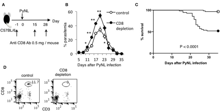
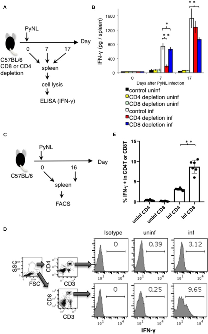
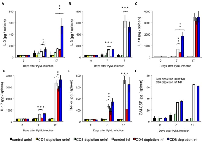
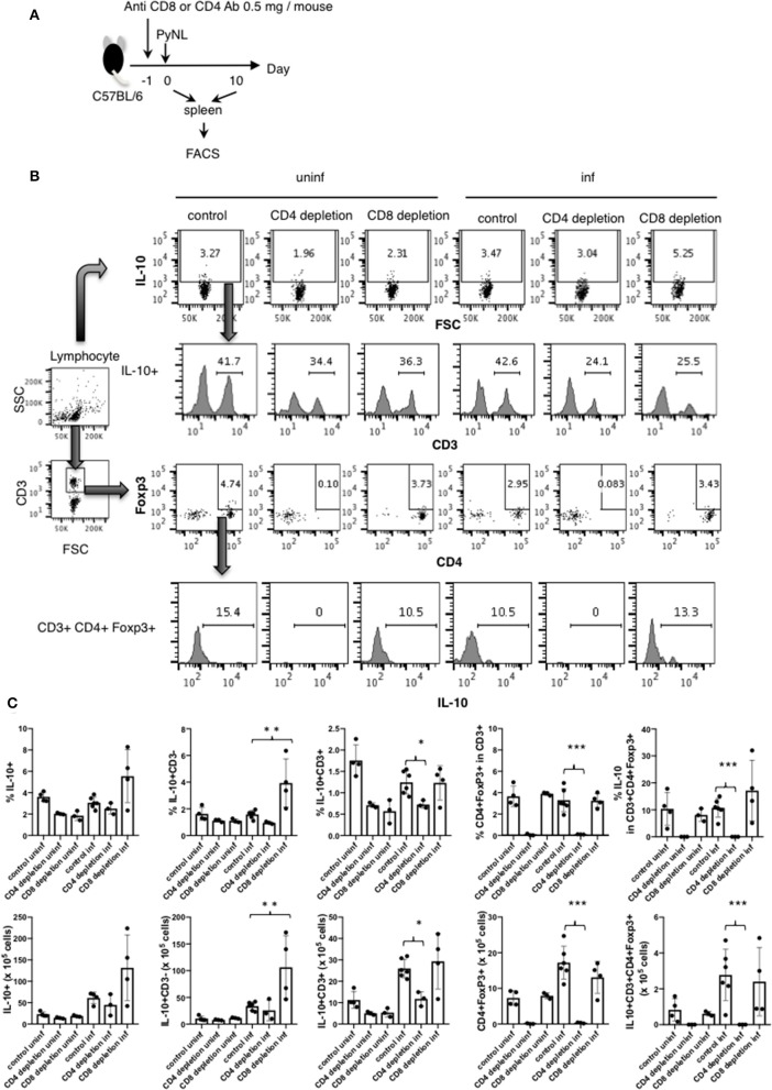
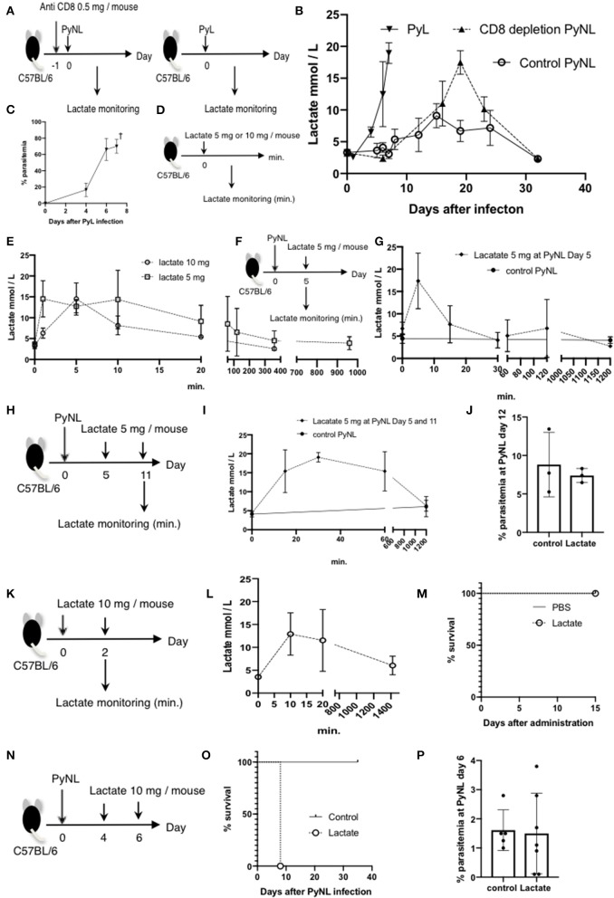
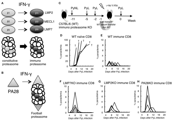
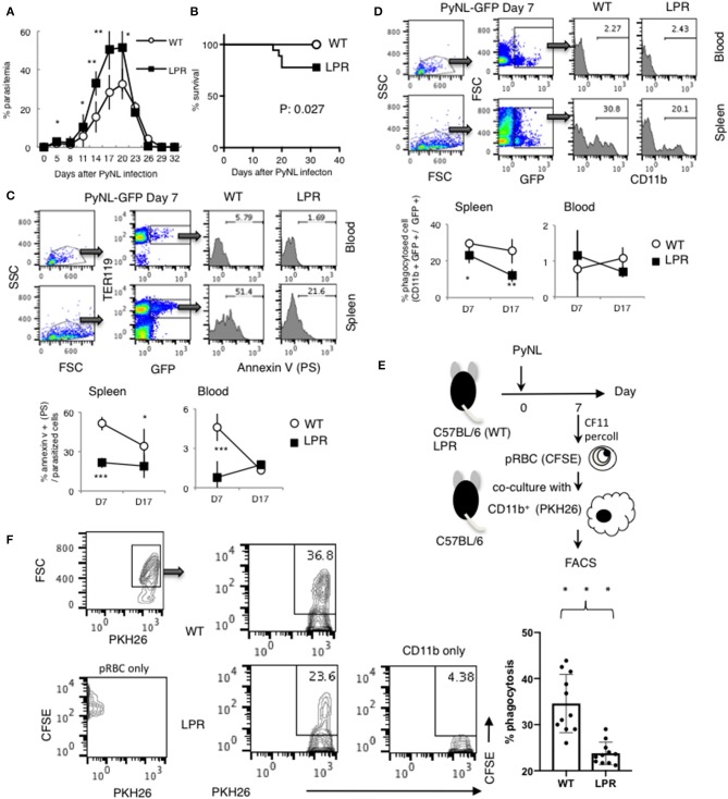
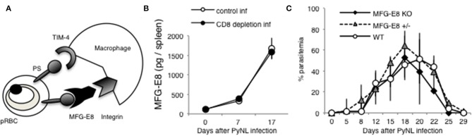
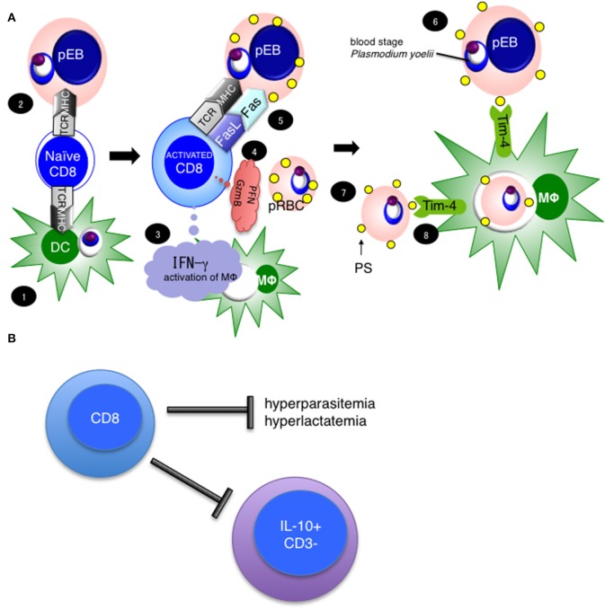
Our previous studies of protective immunity and pathology against blood stage malaria parasites have shown that not only CD4+ T cells, but also CD8+ T cells and macrophages, are important for host defense against blood stage malaria infection. Furthermore, we found that Plasmodium yoelii 17XNL (PyNL) parasitizes erythroblasts, the red blood cell (RBC) precursor cells, which then express MHC class I molecules. In the present study, we analyzed spleen cytokine production. In CD8+ T cell-depleted mice, IL-10 production in early stage infection was increased over two-fold relative to infected control animals and IL-10+ CD3- cells were increased, whereas IFN-γ production in the late stage of infection was decreased. At day 16 after PyNL infection, CD8+ T cells produced more IFN-γ than CD4+ T cells. We evaluated the involvement of the immunoproteasome in induction of immune CD8+ T cells, and the role of Fas in protection against PyNL both of which are downstream of IFN-γ. In cell transfer experiments, at least the single molecules LMP7, LMP2, and PA28 are not essential for CD8+ T cell induction. The Fas mutant LPR mouse was weaker in resistance to PyNL infection than WT mice, and 20% of the animals died. LPR-derived parasitized erythroid cells exhibited less externalization of phosphatidylserine (PS), and phagocytosis by macrophages was impaired. Furthermore, we tried to identify the cause of death in malaria infection. Blood lactate concentration was increased in the CD8+ T cell-depleted PyNL-infected group at day 19 (around peak parasitemia) to similar levels as day 7 after infection with a lethal strain of Py. When we injected mice with lactate at day 4 and 6 of PyNL infection, all mice died at day 8 despite demonstrating low parasitemia, suggesting that hyperlactatemia is one of the causes of death in CD8+ T cell-depleted PyNL-infected mice. We conclude that CD8+ T cells might control cytokine production to some extent and regulate hyperparasitemia and hyperlactatemia in protection against blood stage malaria parasites.

Keywords: CD4 T cell; CD8 T cell; T cell; erythroblast; macrophage; malaria.


Figures

Figure 1
Figure 1
CD8+ T cells contribute to protection against PyNL infection. (A) C57BL/6 mice were injected with anti CD8 Ab or control Ab (0.5 mg/mouse) 1 day before and 15, 28 days after PyNL infection. (B) Parasitemia and (C) survival rate after PyNL infection. Data were pooled from 11 independent experiments [control group: N = 60, white circle; anti-CD8 (CD8+ T cell depletion): N = 56, black circle]. P < 0.0001. Parasitemia values are expressed average ±SD. (D) Depletion of CD8+ T (CD8+ CD3+) cells was confirmed by FACS. *P < 0.05, **P < 0.01.
Figure 2
Figure 2
IFN-γ production from CD4+ or CD8+ T cells from PyNL infected mice and from CD4+ or CD8+ T cell-depleted mice ex vivo. (A) CD4+ or CD8+ T cells were depleted with antibody administration at 1 day before and 15 days after PyNL infection. Spleens were collected from day 0, 7, and 17 after infection. Cells were lysed and centrifuged to remove cell debris, and supernatants were subjected to ELISA (IFN-γ). (B) One representative result from four independent experiments is shown, and is expressed as average ± SD. N = 3–6. (C) C57BL/6 mice were infected with PyNL at day 0. Spleens were collected at day 0 and day 16 after PyNL infection. CD4+ or CD8+ T cells were analyzed by FACS with intracellular IFN-γ staining. (D) Gating strategy is shown in right panel, WBC gated population were further gated with CD4+ and CD3+ or CD8+ and CD3+. Staining of IFN-γ is shown in the histogram. Numbers indicate percentage of IFN-γ staining positive cells in CD4+ T cells or CD8+ T cells. Representative data from two independent experiments are shown. Numbers indicate percentage of IFN-γ staining positive cells in CD4+ T cells or CD8+ T cells. (E) Data are expressed as average ± SD. N = 6. *P < 0.05, **P < 0.01.
Figure 3
Figure 3
Cytokine fluctuations in spleens from CD4+ or CD8+ T cell-depleted mice ex vivo. CD4+ or CD8+ T cells were depleted with antibody administration 1 day before and 15 days after PyNL infection. Spleens were collected on days 0, 7, and 17 after infection. Cells were lysed and centrifuged to remove cell debris, and supernatants were subjected to ELISA (A: IL-2, B: IL-3, C: IL-10, D: IL-17, E: TNF-α, F: GM-CSF). (E) One representative result from four independent experiments is shown, and data are expressed as average ± SD. N = 3–6. **P < 0.01, ***P < 0.001.
Figure 4
Figure 4
IL-10 producing CD3 cells were increased in CD8+ T cell-depleted PyNL-infected mice. (A) C57BL/6 mice were injected with anti-CD8 Ab, anti-CD4 or control Ab (0.5 mg/mouse) 1 day before PyNL infection. Spleens were collected on days 0 or 10 after infection and single cells were stained with fluorescence-conjugated anti-CD3, -CD4, -CD8, -Foxp3, and -IL-10 then analyzed by FACS. (B) Gating strategy is shown in right panel. Lymphocytes were gated and IL-10 staining is shown in top panel. Numbers indicate IL-10 positive cells. IL-10+ cells were further expanded by CD3 (2nd line from top), numbers indicate percentage of CD3+ cells in the IL-10+ population. The lymphocyte gated population was gated with CD3 and then expanded for Foxp3 and CD4, numbers indicate Foxp3+ CD4+ cells in the CD3+ population (3rd line from top). IL-10 histogram of gated cells and number indicate percentage of IL-10+ cells in the CD3+ CD4+ Foxp3+ population (bottom panel). (C) Percentage or number of indicated cells are shown as average ± SD. N = 3–6. *P < 0.05, **P < 0.01, ***P < 0.001.
Figure 5
Figure 5
Lactate is one of the causes of death in CD8+ T cell-depleted PyNL-infected mice. (A) C57BL/6 mice were injected with anti-CD8 Ab or control Ab (0.5 mg/mouse) 1 day before PyNL infection and blood lactate was measured (right panel). C57BL/6 mice were infected with PyL and blood lactate was measured (left panel). (B) Fluctuations in blood lactate in PyL-infected (N = 7), CD8+ T cell-depleted PyNL-infected (N = 3), and control PyNL-infected mice (N = 17) are shown. Data are pooled from three independent experiments. (C) Changes in parasitemia of PyL infected mice are shown. All mice died at day 8 p.i. (D,E) C57BL/6 mice were administered 5 or 10 mg of lactate and blood lactate was monitored. N = 3. (F,G) C57BL/6 mice were infected with PyNL, 5 mg of lactate was administrated on day 5 p.i. and blood lactate was monitored. Control PyNL: N = 6, lactate 5 mg PyNL: N = 3. (H,I) C57BL/6 mice were infected with PyNL and 5 mg of lactate was administered on day 5 and 11 p.i. and blood lactate was monitored. Control PyNL: N = 6, lactate 5 mg PyNL: N = 3. (J) Percentages of parasitemia of mice from experiment H on day 12 p.i. are shown. (K,L) C57BL/6 mice were administered 10 mg of lactate on day 0 and 2 and blood lactate was monitored. N = 3. (M) Survival rate of mice from experiment K is shown. (N) C57BL/6 mice were infected with PyNL and 10 mg of lactate was administered on day 4 and 6. (O) Survival rate and (P) percentage of parasitemia of mice from experiment N are shown. Control: N = 5; 10 mg lactate: N = 7.
Figure 6
Figure 6
Evaluation of immunoproteasome in CD8+ T cell induction in live vaccine model. (A) IFN-γ converts subunits β1, 2, and 5 from the constitutive proteasome into β1i (LMP2), β2i (MECL1), and β5i (LMP7) to construct the immunoproteasome. (B) Proteasomal cap structure PA700 is converted to PA28 by IFN-γ, and PA28 binds to the immunoproteasome to construct a football-like proteasome. (C) Live vaccine model against PyL infection. Wild type (WT) and various KO mice were inoculated with PyNL and PyL infected RBCs at the indicated time points, and CD8+ T cells (1 × 10⁷ cells) from immunized WT donor mice or naïve WT mice were transferred into irradiated (5.5 Gy) WT recipient mice, followed by challenge with PyL infection in recipient mice. Parasitemia of recipient mice receiving donor immune CD8+ T cells from (D): WT naïve, (E): WT infected, (F): LMP7−/−, (G): LMP2−/−, (H): PA28−/−. Each line represents a value from an individual mouse (N = 4–6). Dagger symbols indicate death. Similar results were obtained from two independent experiments.
Figure 7
Figure 7
Fas contributes to protection against PyNL infection. (A) Parasitemia and (B) survival rate are shown. Parasitemia values are expressed as average ± SD. Data are representative of two pooled independent experiments (WT: N = 19, LPR: N = 14). (C) PyNL-GFP was inoculated on day 0, and spleen and peripheral blood were collected at day 7 and 17 after infection for analysis of PS externalization using annexin V and Ter119 (erythroid cell marker) and GFP flow cytometric analysis. One example of analysis in PyNL-GFP at day 7 is shown (Top panel). WBC gated population with dot plot of FSC and SSC were further gated for TER119+ GFP+ infected erythroid cells and expand with Annexin V (PS). Numbers indicate percentage of PS+ cells in infected erythroid cells in blood or spleen. Data are expressed as average ± SD from two pooled independent experiments (bottom panel, WT: N = 11, LPR: N = 9). (D) Percent phagocytosed cells (proportion of CD11b+ GFP+ in GFP+ cells) was determined by flow cytometry. One example of analysis in PyNL-GFP day 7 is shown (Top panel). WBC gated population with dot plot of FSC and SSC were further gated for GFP+ cells which include infected cells and phagocytosed cells and expand with CD11b. Numbers indicate percentage of CD11b+ cells in GFP+ cells in blood or spleen. Data are expressed as average ± SD from two pooled independent experiments (bottom panel, WT: N = 11, LPR: N = 9). (E) Parasitized RBCs (pRBCs) were isolated from peripheral blood 7 days after infection and labeled with CFSE for the phagocytosis assay. CD11b+ macrophages isolated from uninfected mice were labeled with PKH26, co-cultured with pRBCs and analyzed by flow cytometry to determine the phagocytosis rate. (F) One representative result (left panel) and individual data are expressed as % phagocytosis = PKH+ CFSE+ / PKH+ (WT: N = 11, LPR: N = 11) from three pooled independent experiments. *P < 0.05, **P < 0.01, ***P < 0.001.
Figure 8
Figure 8
MFG-E8 is not critical for host defense against PyNL infection. (A) PS is externalized on some pRBCs, and recognized by TIM-4 and MFG-E8. MFG-E8 binds with macrophage integrin. (B) Spleens were collected from day 0, 7, and 17 after infection. Cells were lysed and centrifuged to remove cell debris, and supernatant was subjected to ELISA (MFG-E8). Data were obtained from two pooled independent experiments, and are expressed as average ± SD. N = 6. (C) MFG-E8-KO (-/- and +/- mice) mice were infected with PyNL. Parasitemia is shown (MFG-E8−/−: N = 7, MFG-E8+/−: N = 6, WT: N = 7). Data were obtained from two pooled independent experiments.
Figure 9
Figure 9
Hypothesis and importance of CD8+ T cell mediated protection against blood stage malaria. (A) Our current hypothesis is based on this study and three previously published papers, cited in the main text. (1) CD8+ T cells are activated with antigen cross-presentation from DC or (2) direct antigen presentation from parasitized erythroblasts, resulting in CD8+ T cell IFN-γ Perforin (PFN) and Granzyme B (GzmB) production. (3) IFN-γ activates macrophages. (4) Meanwhile, CD8+ T cells recognize MHC class I on parasitized erythroblasts and stimulate through FasL to (5) Fas on erythroblasts, then (6) induce the externalization of phosphatidylserine (PS) to the surfaces of parasitized erythroblasts. (7) The externalization of PS is a feature of apoptotic cells, and (8) leads to phagocytosis by macrophages. Tim-4 is involved in this process as a PS receptor. (B) CD8+ T cells regulate hyperparasitemia, hyperlactatemia and the number of IL-10+ CD3 cells to protect against PyNL infection.

Similar articles

Cited by

References

    1. Wizel B, Houghten RA, Parker KC, Coligan JE, Church P, Gordon DM, et al. . Irradiated sporozoite vaccine induces HLA-B8-restricted cytotoxic T lymphocyte responses against two overlapping epitopes of the Plasmodium falciparum sporozoite surface protein 2. J Exp Med. (1995) 182:1435–45. 10.1084/jem.182.5.1435 - DOI - PMC - PubMed
    1. Good MF, Doolan DL. Immune effector mechanisms in malaria. Curr Opin Immunol. (1999) 11:412–9. 10.1016/S0952-7915(99)80069-7 - DOI - PubMed
    1. Hafalla JC, Cockburn IA, Zavala F. Protective and pathogenic roles of CD8+ T cells during malaria infection. Parasite Immunol. (2006) 28:15–24. 10.1111/j.1365-3024.2006.00777.x - DOI - PubMed
    1. Marsh K, Kinyanjui S. Immune effector mechanisms in malaria. Parasite Immunol. (2006) 28:51–60. 10.1111/j.1365-3024.2006.00808.x - DOI - PubMed
    1. Kimura K, Kimura D, Matsushima Y, Miyakoda M, Honma K, Yuda M, et al. . CD8+ T cells specific for a malaria cytoplasmic antigen form clusters around infected hepatocytes and are protective at the liver stage of infection. Infect Immun. (2013) 81:3825–34. 10.1128/IAI.00570-13 - DOI - PMC - PubMed

Publication types