

Case Report: Schwannoma of the sigmoid colon: a case report of a rare colonic neoplasm and review of literature
- PMID: 31608147
- PMCID: PMC6777014
- DOI: 10.12688/f1000research.19110.1
Case Report: Schwannoma of the sigmoid colon: a case report of a rare colonic neoplasm and review of literature
Abstract
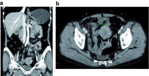
Background: Schwannomas are tumors originating in Schwann cells of the peripheral nerve system and uncommonly develop in the gastrointestinal tract. Sigmoid colon schwannomas are very rare and only 28 cases have been reported. This study aims to report a case of a sigmoid colon schwannoma and present a literature review. Case report: We report a case of a 66-year-old female with asymptomatic sigmoid colon schwannoma. The patient underwent a screening colonoscopy and about 4cm sized submucosal tumor was identified at the sigmoid colon. A colonoscopic biopsy was performed and the microscopic exam revealed an ulcerated lesion with a proliferation of fibroblast-like spindle cells beneath ulcer, which was insufficient for diagnosis. Abdominopelvic computerized tomography (CT) scan showed a well-defined, well-enhancing, round shaped and slightly heterogenous mass at the sigmoid colon. No distant metastasis was identified in abdominopelvic CT and chest CT scans. Carcinoembryonic antigen level was within a normal range (1.33ng/mL). The patient underwent laparoscopic anterior resection. Immunohistochemical staining of the resected specimen showed positivity for S-100 protein in tumor cells and schwannoma was diagnosed post-surgically. Surgical resection margins were free from tumor and no regional lymph node metastasis was reported. Conclusion: Colon schwannomas are rare diseases. Most cases of colon schwannomas are accidentally identified during screening colonoscopy. The tumors usually present as submucosal masses and colonoscopic biopsies are mostly non-diagnostic. Surgical resection is required, and definitive diagnosis is made by confirming S-100 positive tumor cells in immunohistochemical analysis. Most cases are benign; a few cases have been reported to be malignant. Surgical resection with free negative margins is the treatment of choice.
Keywords: Colon; colectomy; colonic neoplasms; schwannoma.
Copyright: © 2019 Kim G et al.
Conflict of interest statement
No competing interests were disclosed.
Figures


References
-
- Uhr A, Singh AP, Munoz J, et al. : Colonic Schwannoma: A Case Study and Literature Review of a Rare Entity and Diagnostic Dilemma. Am Surg. 2016;82(12):1183–86. - PubMed
Publication types
MeSH terms
LinkOut - more resources
Full Text Sources