

Traumatic microbleeds suggest vascular injury and predict disability in traumatic brain injury
- PMID: 31608359
- PMCID: PMC6821371
- DOI: 10.1093/brain/awz290
Traumatic microbleeds suggest vascular injury and predict disability in traumatic brain injury
Abstract
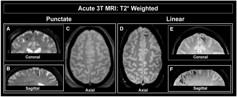
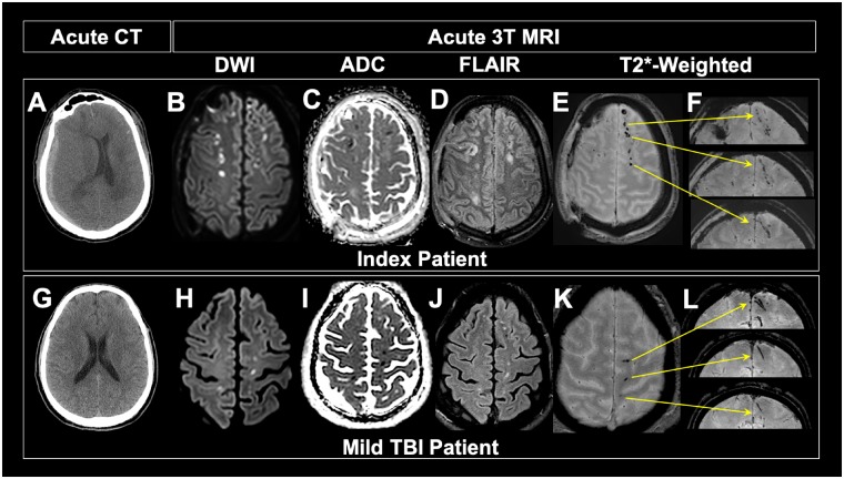
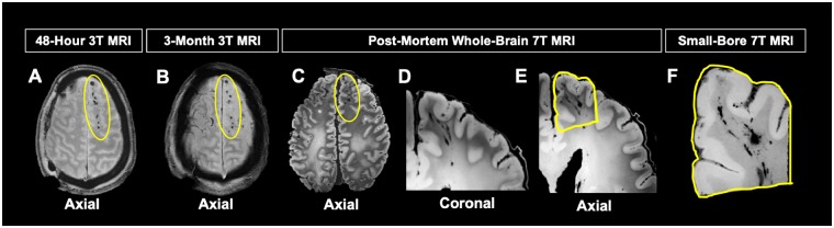
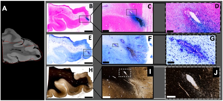
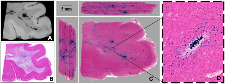
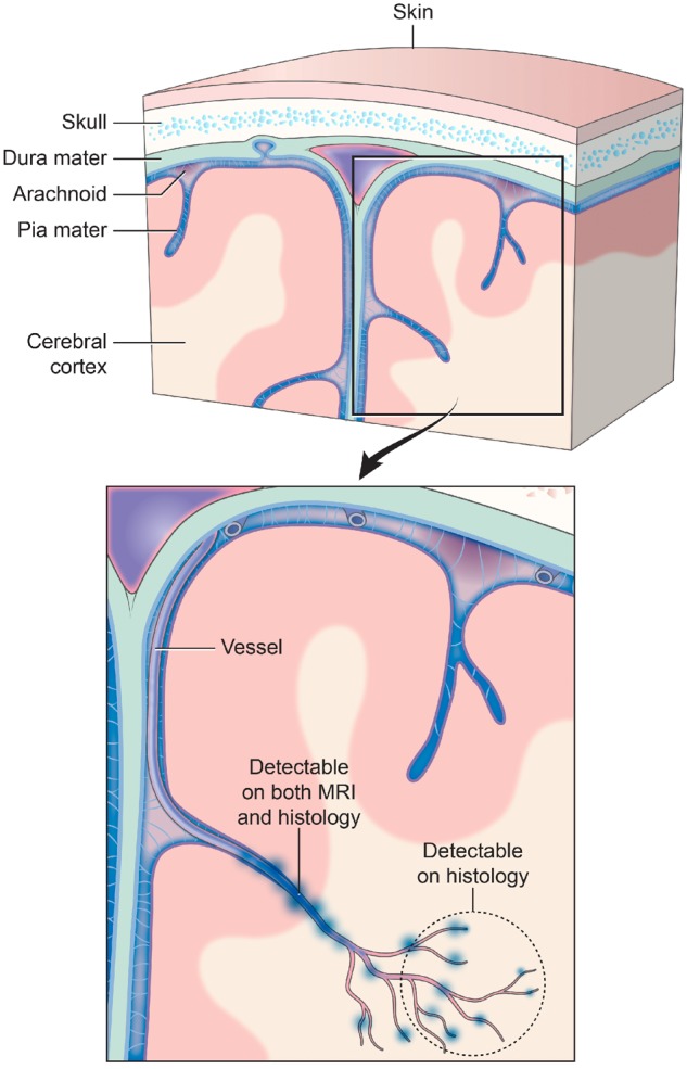
Traumatic microbleeds are small foci of hypointensity seen on T2*-weighted MRI in patients following head trauma that have previously been considered a marker of axonal injury. The linear appearance and location of some traumatic microbleeds suggests a vascular origin. The aims of this study were to: (i) identify and characterize traumatic microbleeds in patients with acute traumatic brain injury; (ii) determine whether appearance of traumatic microbleeds predict clinical outcome; and (iii) describe the pathology underlying traumatic microbleeds in an index patient. Patients presenting to the emergency department following acute head trauma who received a head CT were enrolled within 48 h of injury and received a research MRI. Disability was defined using Glasgow Outcome Scale-Extended ≤6 at follow-up. All magnetic resonance images were interpreted prospectively and were used for subsequent analysis of traumatic microbleeds. Lesions on T2* MRI were stratified based on 'linear' streak-like or 'punctate' petechial-appearing traumatic microbleeds. The brain of an enrolled subject imaged acutely was procured following death for evaluation of traumatic microbleeds using MRI targeted pathology methods. Of the 439 patients enrolled over 78 months, 31% (134/439) had evidence of punctate and/or linear traumatic microbleeds on MRI. Severity of injury, mechanism of injury, and CT findings were associated with traumatic microbleeds on MRI. The presence of traumatic microbleeds was an independent predictor of disability (P < 0.05; odds ratio = 2.5). No differences were found between patients with punctate versus linear appearing microbleeds. Post-mortem imaging and histology revealed traumatic microbleed co-localization with iron-laden macrophages, predominately seen in perivascular space. Evidence of axonal injury was not observed in co-localized histopathological sections. Traumatic microbleeds were prevalent in the population studied and predictive of worse outcome. The source of traumatic microbleed signal on MRI appeared to be iron-laden macrophages in the perivascular space tracking a network of injured vessels. While axonal injury in association with traumatic microbleeds cannot be excluded, recognizing traumatic microbleeds as a form of traumatic vascular injury may aid in identifying patients who could benefit from new therapies targeting the injured vasculature and secondary injury to parenchyma.
Keywords: MRI biomarkers of traumatic brain injury; mild traumatic brain injury; radiological-pathological analysis; traumatic microbleeds; traumatic vascular injury.
Published by Oxford University Press on behalf of the Guarantors of Brain 2019. This work is written by US Government employees and is in the public domain in the US.
Figures
References
-
- Adams JH, Graham DI, Murray LS, Scott G. Diffuse axonal injury due to nonmissle head injury in humans: an analysis of 45 cases. Ann Neurol 1982; 12: 557–63. - PubMed
-
- Babikian T, Freier MC, Tong KA, Nickerson JP, Wall CJ, Holshouser BA, et al.Susceptibility weighted imaging: neuropsychologic outcome and pediatric head injury. Pediatr Neurol 2005; 33: 184–94. - PubMed
-
- Beauchamp MH, Beare R, Ditchfield M, Coleman L, Babl FE, Kean M, et al.Susceptibility weighted imaging and its relationship to outcome after pediatric traumatic brain injury. Cortex 2013; 49: 591–8. - PubMed
-
- Colbert CA, Holshouser BA, Aaen GS, Sheridan C, Oyoyo U, Kido D, et al.Value of cerebral microhemorrhages detected with susceptibility-weighted MR imaging for prediction of long-term outcome in children with nonaccidental trauma. Radiology 2010; 256: 898–905. - PubMed
Publication types
MeSH terms
Substances
LinkOut - more resources
Full Text Sources
Medical
