

Ginsenoside Rb1 Prevents Dysfunction of Endothelial Cells by Suppressing Inflammatory Response and Apoptosis in the High-Fat Diet Plus Balloon Catheter-Injured Rabbit Model via the G Protein-Coupled Estrogen Receptor-Mediated Phosphatidylinositol 3-Kinases (PI3K)/Akt Pathway
- PMID: 31609302
- PMCID: PMC6791186
- DOI: 10.12659/MSM.912986
Ginsenoside Rb1 Prevents Dysfunction of Endothelial Cells by Suppressing Inflammatory Response and Apoptosis in the High-Fat Diet Plus Balloon Catheter-Injured Rabbit Model via the G Protein-Coupled Estrogen Receptor-Mediated Phosphatidylinositol 3-Kinases (PI3K)/Akt Pathway
Abstract
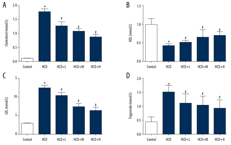
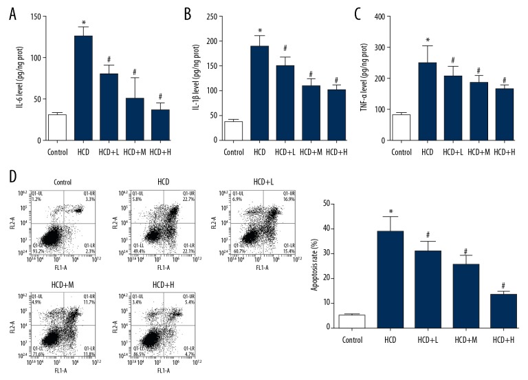
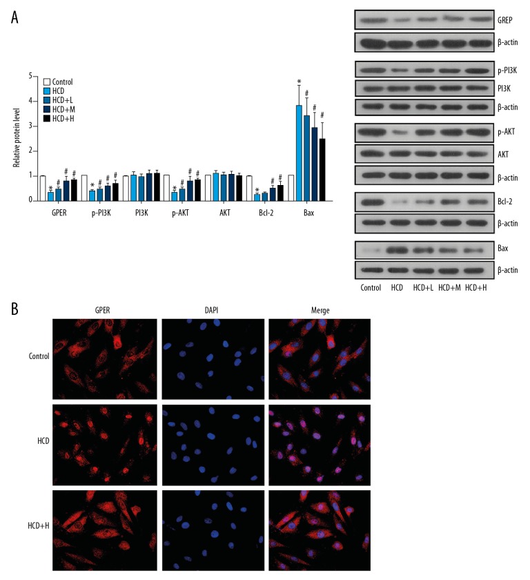
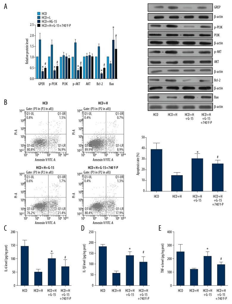
<strong>BACKGROUND</strong> The initiation of atherosclerosis (AS) is attributed to the dysfunction of endothelial cells (ECs) via the inhibition of g protein-coupled estrogen receptor (GPER). In the current study, we assessed the potential of Ginsenoside Rb1 (Rb1) to attenuate the dysfunction of ECs via GPER-mediated PI3K/Akt pathway. <strong>MATERIAL AND METHODS</strong> AS was induced in rabbits and then the AS rabbits were treated with Rb1. Thereafter, the ECs were isolated from AS and healthy rabbits, and treated with Rb1. The effect of Rb1 on blood lipid levels in AS rabbits and on apoptosis, inflammatory response, and GPER/PI3K/Akt axis activity in ECs was detected. Furthermore, the activities of GPER and PI3K were modulated to verify the key role of the axis in the anti-AS effect of Rb1. <strong>RESULTS</strong> The levels of total cholesterol, low-density lipoprotein (LDL), and triglyceride in AS rabbits were suppressed by Rb1 while the high-density lipoprotein (HDL) level was increased. In in vitro assays, Rb1 administration inhibited apoptosis process and the production of pro-inflammation cytokines in AS ECs. The expression levels of GPER, p-PI3K, and p-Akt were upregulated by Rb1, associated with the increased level of Bcl-2 and reduced level of Bax. When the activity of GPER was inhibited by GP-15 in AS ECs, the treatment effect of Rb1 was blocked. However, the activation of PI3K could restore the protective effect of Rb1 after the inhibition of GPER. <strong>CONCLUSIONS</strong> The anti-AS potential of Rb1 was exerted by restoring the regular function of ECs via the activation of GPER-mediated PI3K/Akt signaling.
Conflict of interest statement
None.
Figures
References
-
- Furchgott RF. Introduction to EDRF research. J Cardiovasc Pharma. 1993;22(Suppl 7):S1–2. - PubMed
-
- Galili O, Versari D, Sattler KJ, et al. Early experimental obesity is associated with coronary endothelial dysfunction and oxidative stress. Am J Physiol-Heart C. 2007;292:H904. - PubMed
-
- Barton M, Haudenschild CC. Endothelium and atherogenesis: Endothelial therapy revisited. J Cardiovasc Pharma. 2001;38(Suppl 2):S23. - PubMed
-
- Traupe T, Ortmann J, Münter K, Barton M. Endothelial therapy of atherosclerosis and its risk factors. Curr Vasc Pharmacol. 2003;1:111–21. - PubMed
MeSH terms
Substances
LinkOut - more resources
Full Text Sources
Medical
Research Materials
Miscellaneous
