

The Appendix in Parkinson's Disease: From Vestigial Remnant to Vital Organ?
- PMID: 31609697
- PMCID: PMC6839473
- DOI: 10.3233/JPD-191703
The Appendix in Parkinson's Disease: From Vestigial Remnant to Vital Organ?
Abstract
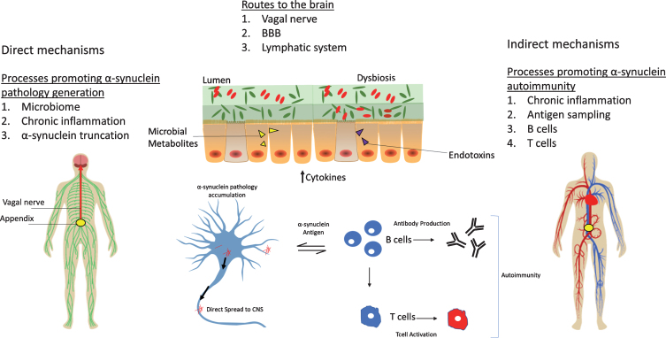
Parkinson's disease (PD) has long been considered a brain disease, but studies now point to the gastrointestinal (GI) tract as a potential starting point for PD. In particular, the human vermiform appendix has been implicated in PD. The appendix is a tissue rich in immune cells, serving as part of the gut-associated lymphoid tissue and as a storehouse for the gut microbiome. The functions of the appendix converge with recent evidence demonstrating that gut inflammation and shifts in the microbiome are linked to PD. Some epidemiological studies have linked removal of the appendix to lowered PD risk, though there is controversy among these associations. What is apparent is that there is an abundance of aggregated forms of α-synuclein in the appendix relevant to PD pathology. α-Synuclein pathology is thought to propagate from gut to brain via the vagus nerve, which innervates GI tract locations, including the appendix. Remarkably, α-synuclein aggregates in the appendix occur not only in PD patients, but are also present in healthy individuals. This has led to the proposal that in the appendix α-synuclein aggregates are not unique to PD. Moreover, the molecular events leading to PD and the mechanisms by which α-synuclein aggregates transfers from gut to brain may be identifiable in the human appendix. The influence of the appendix on GI inflammation, autoimmunity, microbiome storage, and the lymphatic system may be yet unexplored mechanisms by which the appendix contributes to PD. Overall, the appendix represents a promising tissue site to advance our understanding of PD pathobiology.
Keywords: Parkinson’s disease; appendix; gastrointestinal tract; vagus nerve; α-Synuclein.
Conflict of interest statement
The authors have no conflict of interest to report.
Figures
References
-
- Smith HF, P W, SH K, M L (2017) Morphological evolution of the mammalian cecum and cecal appendix. Comptes Rendus Palevol 16, 39–57.
-
- Vitetta L, Chen J, Clarke S (2019) The vermiform appendix: An immunological organ sustaining a microbiome inoculum. Clin Sci (Lond) 133, 1–8. - PubMed
-
- Palestrant D, Holzknecht ZE, Collins BH, Parker W, Miller SE, Bollinger RR (2004) Microbial biofilms in the gut: Visualization by electron microscopy and by acridine orange staining. Ultrastruct Pathol 28, 23–27. - PubMed
-
- Masahata K, Umemoto E, Kayama H, Kotani M, Nakamura S, Kurakawa T, Kikuta J, Gotoh K, Motooka D, Sato S, Higuchi T, Baba Y, Kurosaki T, Kinoshita M, Shimada Y, Kimura T, Okumura R, Takeda A, Tajima M, Yoshie O, Fukuzawa M, Kiyono H, Fagarasan S, Iida T, Ishii M, Takeda K (2014) Generation of colonic IgA-secreting cells in the caecal patch. Nat Commun 5, 3704. - PubMed
Publication types
MeSH terms
Substances
Grants and funding
LinkOut - more resources
Full Text Sources
Medical
