. 2019 Oct 14;17(10):e3000468.
doi: 10.1371/journal.pbio.3000468. eCollection 2019 Oct.

KHDC3L mutation causes recurrent pregnancy loss by inducing genomic instability of human early embryonic cells

Affiliations

KHDC3L mutation causes recurrent pregnancy loss by inducing genomic instability of human early embryonic cells

Weidao Zhang et al. PLoS Biol. .

Abstract

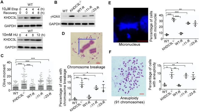
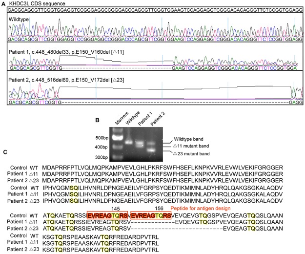
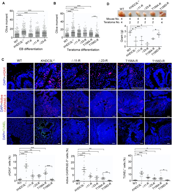
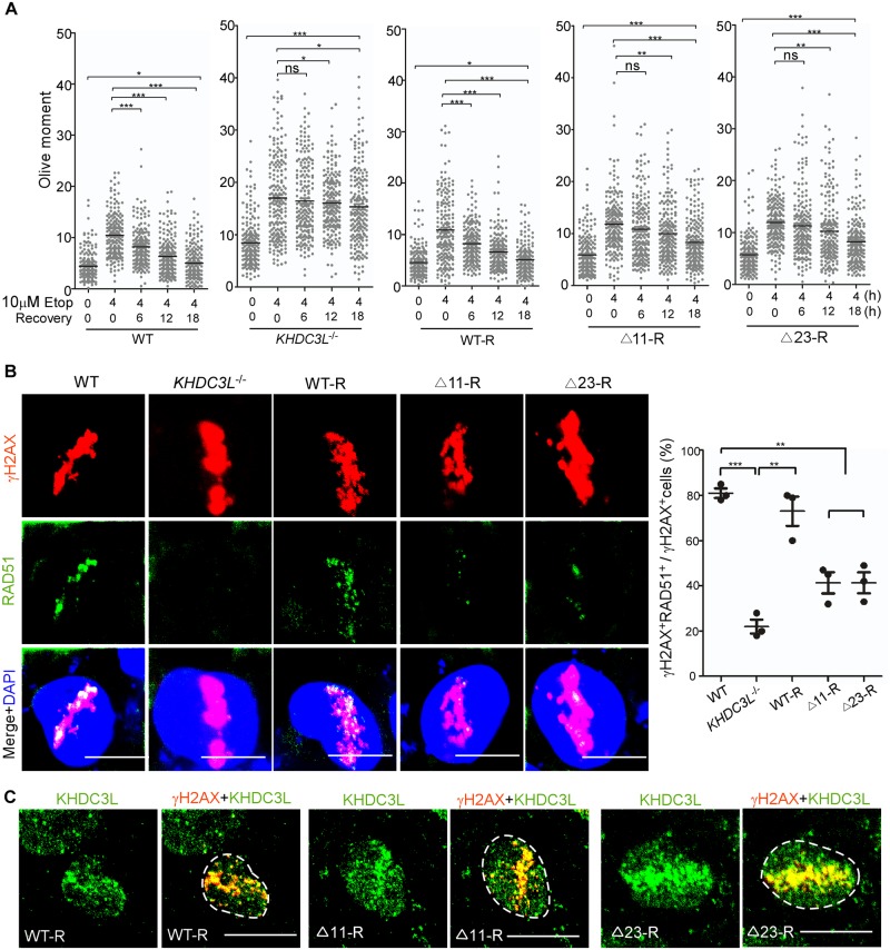
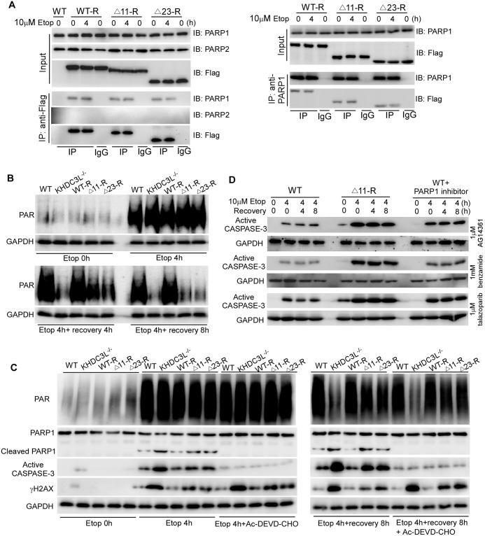
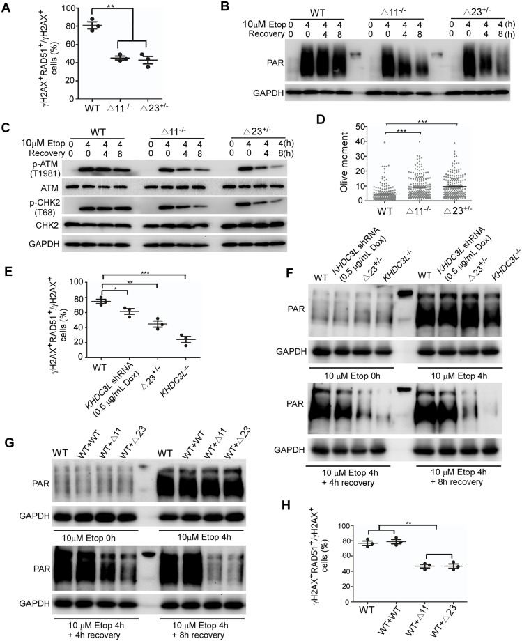
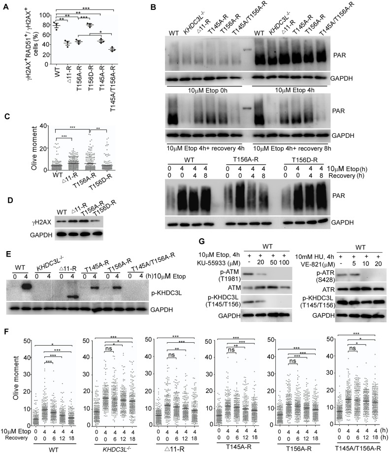
Recurrent pregnancy loss (RPL) is an important complication in reproductive health. About 50% of RPL cases are unexplained, and understanding the genetic basis is essential for its diagnosis and prognosis. Herein, we report causal KH domain containing 3 like (KHDC3L) mutations in RPL. KHDC3L is expressed in human epiblast cells and ensures their genome stability and viability. Mechanistically, KHDC3L binds to poly(ADP-ribose) polymerase 1 (PARP1) to stimulate its activity. In response to DNA damage, KHDC3L also localizes to DNA damage sites and facilitates homologous recombination (HR)-mediated DNA repair. KHDC3L dysfunction causes PARP1 inhibition and HR repair deficiency, which is synthetically lethal. Notably, we identified two critical residues, Thr145 and Thr156, whose phosphorylation by Ataxia-telangiectasia mutated (ATM) is essential for KHDC3L's functions. Importantly, two deletions of KHDC3L (p.E150_V160del and p.E150_V172del) were detected in female RPL patients, both of which harbor a common loss of Thr156 and are impaired in PARP1 activation and HR repair. In summary, our study reveals both KHDC3L as a new RPL risk gene and its critical function in DNA damage repair pathways.


Conflict of interest statement

The authors have declared that no competing interests exist.

Figures

Fig 1
Fig 1. KHDC3L preserves genomic stability of hESCs.
(A) KHDC3L protein expression was up-regulated by the treatments of Etop (upper panel) and HU (lower panel) in hESCs. Immunoblotting with γH2AX (B) and neutral comet assay (C) (n = 200 from two independent replicates) revealed that knockout of KHDC3L (KHDC3L−/−) in hESCs led to elevated DNA DSBs. This defect was fully rescued by reexpression of WT KHDC3L (WT-R) but not mutant proteins Δ11 or Δ23 (Δ11-R, Δ23-R) identified in patients with RPL. hESCs with dysfunctional KHDC3L (KHDC3L−/−, Δ11-R, Δ23-R) displayed a higher level of chromosome breaks (D), micronuclei (E), and aneuploidy (F) than those with functional KHDC3L (WT and WT-R) (n = 50 in one replicate, and total three independent replicates in D-F). Data represent mean ± SEM. *p < 0.05, **p < 0.01, ***p < 0.001. Scale bars, 10 μm. Underlying numerical values in (C-F) can be found in S1 Data. Δ11, p.E150_V160del; Δ23, p.E150_V172del; DSB, double-strand break; Etop, etoposide; GAPDH, glyceraldehyde 3-phosphate dehydrogenase; hESC, human embryonic stem cell; HU, hydroxyurea; KHDC3L, KH domain containing 3 like; WT, wild type.
Fig 2
Fig 2. KHDC3L is mutated in patients with RPL.
(A) Sanger sequencing identified Δ11 (NM_001017361, c.448-480del33) and Δ23 (NM_001017361, c.448_516del69) heterozygous deletions of KHDC3L in two unrelated patients. (B) PCR amplification confirmed the heterozygous mutations of KHDC3L in two patients. (C) Alignment of protein sequences of WT KHDC3L and two mutants (Δ11 and Δ23) identified in two unrelated patients with RPL. Δ11, p.E150_V160del; Δ23, p.E150_V172del; KHDC3L, KH domain containing 3 like; RPL, recurrent pregnancy loss; WT, wild type.
Fig 3
Fig 3. KHDC3L deficiency causes DNA damage and apoptosis in cells differentiated from hESCs.
hESCs underwent in vitro EB differentiation (A) or in vivo teratoma differentiation (B). Neutral comet assay showed that differentiated progenies from hESCs with deficient KHDC3L (KHDC3L−/−, Δ11-R, and Δ23-R) had severe DNA DSBs (n = 200 from two independent experiments). (C) Immunostaining with γH2AX, active CASPASE-3, and TUNEL validated the higher level of DNA DSBs and apoptosis in teratoma cells differentiated from hESCs without KHDC3L or expressing the mutant KHDC3L (KHDC3L−/−, Δ11-R, Δ23-R, and T156A-R). (D) Fewer and smaller teratomas were formed by hESCs in the absence of functional KHDC3L (KHDC3L−/−, Δ11-R, Δ23-R, and T156A-R) (above, the injected mice numbers and the teratomas numbers; below, quantification of teratomas weight). Student two-tailed t test was performed for statistical analysis. Data represent mean ± SEM. *p < 0.05, **p < 0.01, ***p < 0.001. Scale bars, 100 μm. Underlying numerical values in A-D can be found in S1 Data. Δ11, p.E150_V160del; Δ23, p.E150_V172del; DSB, double-strand break; EB, embryoid body; TUNEL, TdT-mediated dUTP Nick-End Labeling; hESC, human embryonic stem cell; KHDC3L, KH domain containing 3 like; WT, wild type.
Fig 4
Fig 4. KHDC3L deficiency impairs HR-mediated DNA repair in hESCs.
(A) Neutral comet assay revealed that DSB repair was less efficient in hESCs with deficient KHDC3L (KHDC3L−/−, Δ11-R, and Δ23-R) than in hESCs with proficient KHDC3L (WT and WT-R) (n = 200 from two independent experiments). (B) hESCs were laser micro-irradiated and examined following 2 h of recovery. hESCs with deficient KHDC3L (KHDC3L−/−, Δ11-R, and Δ23-R) are impaired in the recruitment of RAD51 to DSB sites labeled with γH2AX, indicating their reduced HR repair capacity (n = 50 in one replicate, total three independent replicates). (C) WT KHDC3L (WT-R) localizes to DSB sites. The Δ11 (Δ11-R) and Δ23 mutations (Δ23-R) did not impair this cellular localization (n = 10 in one replicate, total three independent replicates). Student two-tailed t test was performed for statistical analysis. Data represent mean ± SEM. *p < 0.05, **p < 0.01, ***p < 0.001. Scale bars, 10 μm. Underlying numerical values in (A) and (B) can be found in S1 Data. Δ11, p.E150_V160del; Δ23, p.E150_V172del; DSB, double-strand break; Etop, etoposide; hESC, human embryonic stem cell; HR, homologous recombination; KHDC3L, KH domain containing 3 like; ns, not significant; RAD51, RAS associated with diabetes protein 51; WT, wild type.
Fig 5
Fig 5. KHDC3L deficiency impairs PARP1 activation in hESCs.
(A) Reciprocal coimmunoprecipitation revealed the physical and constitutive interaction of KHDC3L with PARP1 but not PARP2. The Δ11 or Δ23 mutation did not impair this interaction. (B) After Etop treatment, hESCs with proficient KHDC3L (WT and WT-R) maintained high PAR levels, whereas those with deficient KHDC3L (KHDC3L−/−, Δ11-R, and Δ23-R) failed to efficiently sustain PAR levels. (C) Apoptosis inhibitor Ac-DEVD-CHO successfully suppressed apoptosis and PARP1 cleavage. However, it did not affect the levels of PAR and γH2AX. (D) Due to synthetic lethality caused by simultaneous impairment of HR repair and PARP1 activation in hESCs with deficient KHDC3L, Δ11-R hESCs were more sensitive than WT hESCs or WT hESCs treated with the indicated PARP1 inhibitors in response to genotoxic insults. Δ11, p.E150_V160del; Δ23, p.E150_V172del; Etop, etoposide; GAPDH, glyceraldehyde 3-phosphate dehydrogenase; hESC, human embryonic stem cell; HR, homologous recombination; IB, immunoblot; IgG, immunoglobulin G; IP, immunoprecipitation; KHDC3L, KH domain containing 3 like; PAR, poly(ADP-ribose); PARP, PAR polymerase; WT, wild type.
Fig 6
Fig 6. The Δ11 and Δ23 mutants display a dominant-negative effect.
Homozygous deletion of 11aa (Δ11−/−) and heterozygous deletion of 23aa (Δ23+/−) in hESCs caused similar extent of defects in HR-mediated DNA repair (A) (n = 50 in one replicates, total three independent experiments), PARP1 activation (B), and ATM-CHK2 signaling (C). Consequently, Δ11−/− and Δ23+/− hESCs accumulated similar level of DNA DSBs (D) (n = 200 from two independent experiments). Compared to Δ23+/− mutation, partial knockdown of KHDC3L caused milder defects in RAD51 recruitment to DSB sites (E) and PARP1 activation (F). Expression of Δ11 or Δ23 mutants, but not WT KHDC3L, in WT ESCs impaired PARP1 activation (G) and HR-mediated repair (H). Student two-tailed t test was performed for statistical analysis. Data represent mean ± SEM. *p < 0.05, **p < 0.01, ***p < 0.001. Underlying numerical values in (A), (D), (E), and (H) can be found in S1 Data. Δ11, p.E150_V160del; Δ23, p.E150_V172del; aa, amino acid; ATM, Ataxia-telangiectasia mutated; CHK2, checkpoint kinase 2; Dox, doxycycline; DSB, double-strand break; ESC, embryonic stem cell; Etop, etoposide; GAPDH, glyceraldehyde 3-phosphate dehydrogenase; hESC, human ESC; HR, homologous recombination; KHDC3L, KH domain containing 3 like; PAR, poly(ADP-ribose); PARP, PAR polymerase; shRNA, short hairpin RNA; WT, wild type.
Fig 7
Fig 7. Phosphorylation of T156 and T145 sites in KHDC3L by ATM is required for HR repair and PARP1 activation.
(A) T156A, T145A, and Δ11 mutations caused similar level of defect in recruiting RAD51 to DNA DSB sites, whereas T156D had no effects on RAD51 recruitment. Double mutations of T156A/T145A displayed a more severe phenotype than those of individual mutation (n = 50 in one replicate, total three independent experiments). (B) PARP1 activity in hESCs with WT or mutant KHDC3L proteins. Neutral comet assay (C) (n = 200 from two independent experiments) and immunoblotting (D) confirmed that hESCs expressing Δ11 or T156A mutant KHDC3L accumulated a higher level of DNA DSBs than did WT hESCs or ESCs expressing T156D. (E) Phosphorylation of T145/T156 sites at different hESC lines. (F) T145A, T156A, and Δ11 mutations displayed similar level of defects in DSB repair (n = 200 from two independent experiments). Similar to KHDC3L−/−, T156A/T145A double mutations caused more-severe defects. (G) Complete repression of ATM activation by KU-55933 eliminated the phosphorylation of T145/T156 in KHDC3L, whereas inhibition of ATR activity by VE-821 had no effects. Student two-tailed t test was performed for statistical analysis. Data represent mean ± SEM. *p < 0.05, **p < 0.01, ***p < 0.001. Underlying numerical values in (A), (C), and (F) can be found in S1 Data. Δ11, p.E150_V160del; ATM, Ataxia-telangiectasia mutated; ATR, Ataxia-telangiectasia and Rad3-related protein; DSB, double-strand break; ESC, embryonic stem cell; Etop, etoposide; GAPDH, glyceraldehyde 3-phosphate dehydrogenase; hESC, human ESC; HR, homologous recombination; KHDC3L, KH domain containing 3 like; ns, not significant; PAR, poly(ADP-ribose); PARP, PAR polymerase; RAD51, RAS associated with diabetes protein 51; WT, wild type.

Similar articles

Cited by

References

    1. Practice Committee of the American Society for Reproductive M. Evaluation and treatment of recurrent pregnancy loss: a committee opinion. Fertility and sterility. 2012;98(5):1103–11. 10.1016/j.fertnstert.2012.06.048 - DOI - PubMed
    1. Arias-Sosa LA, Acosta ID, Lucena-Quevedo E, Moreno-Ortiz H, Esteban-Perez C, Forero-Castro M. Genetic and epigenetic variations associated with idiopathic recurrent pregnancy loss. Journal of assisted reproduction and genetics. 2018;35(3):355–66. 10.1007/s10815-017-1108-y - DOI - PMC - PubMed
    1. Hyde KJ, Schust DJ. Genetic considerations in recurrent pregnancy loss. Cold Spring Harbor perspectives in medicine. 2015;5(3):a023119 10.1101/cshperspect.a023119 - DOI - PMC - PubMed
    1. Simpson JL. Causes of fetal wastage. Clinical obstetrics and gynecology. 2007;50(1):10–30. 10.1097/GRF.0b013e31802f11f6 - DOI - PubMed
    1. Hassold T, Hunt P. To err (meiotically) is human: the genesis of human aneuploidy. Nature reviews Genetics. 2001;2(4):280–91. 10.1038/35066065 - DOI - PubMed

Publication types

MeSH terms