

CavBench: A benchmark for protein cavity detection methods
- PMID: 31609980
- PMCID: PMC6791542
- DOI: 10.1371/journal.pone.0223596
CavBench: A benchmark for protein cavity detection methods
Abstract
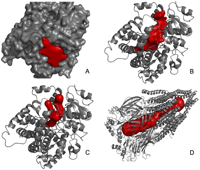
Extensive research has been applied to discover new techniques and methods to model protein-ligand interactions. In particular, considerable efforts focused on identifying candidate binding sites, which quite often are active sites that correspond to protein pockets or cavities. Thus, these cavities play an important role in molecular docking. However, there is no established benchmark to assess the accuracy of new cavity detection methods. In practice, each new technique is evaluated using a small set of proteins with known binding sites as ground-truth. However, studies supported by large datasets of known cavities and/or binding sites and statistical classification (i.e., false positives, false negatives, true positives, and true negatives) would yield much stronger and reliable assessments. To this end, we propose CavBench, a generic and extensible benchmark to compare different cavity detection methods relative to diverse ground truth datasets (e.g., PDBsum) using statistical classification methods.
Conflict of interest statement
The authors have declared that no competing interests exist.
Figures

Similar articles
-
Anatomy of protein pockets and cavities: measurement of binding site geometry and implications for ligand design.Protein Sci. 1998 Sep;7(9):1884-97. doi: 10.1002/pro.5560070905. Protein Sci. 1998. PMID: 9761470 Free PMC article.
-
The scoring bias in reverse docking and the score normalization strategy to improve success rate of target fishing.PLoS One. 2017 Feb 14;12(2):e0171433. doi: 10.1371/journal.pone.0171433. eCollection 2017. PLoS One. 2017. PMID: 28196116 Free PMC article.
-
Roll: a new algorithm for the detection of protein pockets and cavities with a rolling probe sphere.Bioinformatics. 2010 Jan 1;26(1):46-52. doi: 10.1093/bioinformatics/btp599. Epub 2009 Oct 21. Bioinformatics. 2010. PMID: 19846440
-
dxTuber: detecting protein cavities, tunnels and clefts based on protein and solvent dynamics.J Mol Graph Model. 2011 Jun;29(7):895-905. doi: 10.1016/j.jmgm.2011.02.003. Epub 2011 Feb 24. J Mol Graph Model. 2011. PMID: 21420887
-
[Molecular docking: role of intermolecular contacts in formation of complexes of proteins with nucleotides and peptides].Bioorg Khim. 2010 Jul-Aug;36(4):482-92. doi: 10.1134/s1068162010040023. Bioorg Khim. 2010. PMID: 20823916 Review. Russian.
Cited by
-
In Silico Analysis of Peptide Macrocycle -Protein Interactions.Methods Mol Biol. 2022;2371:317-334. doi: 10.1007/978-1-0716-1689-5_17. Methods Mol Biol. 2022. PMID: 34596856
-
PDBspheres: a method for finding 3D similarities in local regions in proteins.NAR Genom Bioinform. 2022 Oct 10;4(4):lqac078. doi: 10.1093/nargab/lqac078. eCollection 2022 Dec. NAR Genom Bioinform. 2022. PMID: 36225529 Free PMC article.
-
A Comprehensive Mapping of the Druggable Cavities within the SARS-CoV-2 Therapeutically Relevant Proteins by Combining Pocket and Docking Searches as Implemented in Pockets 2.0.Int J Mol Sci. 2020 Jul 21;21(14):5152. doi: 10.3390/ijms21145152. Int J Mol Sci. 2020. PMID: 32708196 Free PMC article.
-
pyCAST, a Python package for the detection of cavities on surface proteins.Comput Struct Biotechnol J. 2025 Aug 11;27:3589-3597. doi: 10.1016/j.csbj.2025.07.054. eCollection 2025. Comput Struct Biotechnol J. 2025. PMID: 40821718 Free PMC article.
-
Predicting binding sites from unbound versus bound protein structures.Sci Rep. 2020 Sep 28;10(1):15856. doi: 10.1038/s41598-020-72906-7. Sci Rep. 2020. PMID: 32985584 Free PMC article.
References
-
- Shoichet B, Kuntz I, Bodian D. Molecular docking using shape descriptors. Journal of Computational Chemistry. 1992;13(3):380–397. 10.1002/jcc.540130311 - DOI
Publication types
MeSH terms
Substances
LinkOut - more resources
Full Text Sources
