

Single-dose combination nanovaccine induces both rapid and long-lived protection against pneumonic plague
- PMID: 31610342
- PMCID: PMC7012387
- DOI: 10.1016/j.actbio.2019.10.016
Single-dose combination nanovaccine induces both rapid and long-lived protection against pneumonic plague
Abstract
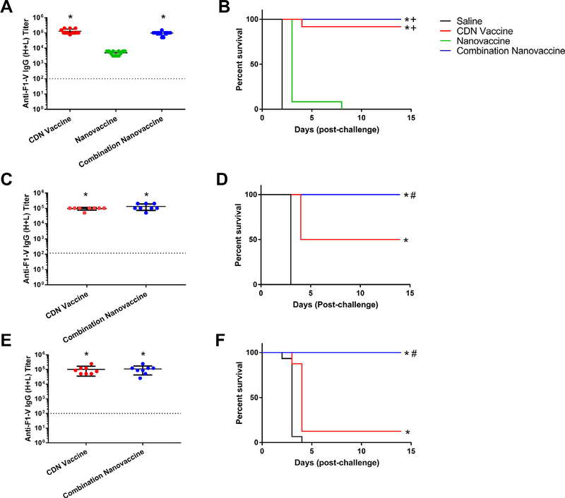
Yersinia pestis, the causative agent of pneumonic plague, induces a highly lethal infection if left untreated. Currently, there is no FDA-approved vaccine against this pathogen; however, USAMRIID has developed a recombinant fusion protein, F1-V, that has been shown to induce protection against pneumonic plague. Many F1-V-based vaccine formulations require prime-boost immunization to achieve protective immunity, and there are limited reports of rapid induction of protective immunity (≤ 14 days post-immunization (DPI)). The STimulator of INterferon Genes agonists cyclic dinucleotides (CDNs) have been shown to be promising vaccine adjuvants. Polyanhydride nanoparticle-based vaccines (i.e., nanovaccines) have also shown to enhance immune responses due to their dual functionality as adjuvants and delivery vehicles. In this work, a combination nanovaccine was designed that comprised F1-V-loaded nanoparticles combined with the CDN, dithio-RP,RP-cyclic di-guanosine monophosphate, to induce rapid and long-lived protective immunity against pneumonic plague. All mice immunized with a single dose combination nanovaccine were protected from Y. pestis lethal challenge within 14 DPI and demonstrated enhanced protection over F1-V adjuvanted with CDNs alone at challenge doses ≥7000 CFU Y. pestis CO92. In addition, 75% of mice receiving the single dose of the combination nanovaccine were protected from challenge at 182 DPI, while maintaining high levels of antigen-specific serum IgG. ELISPOT analysis of vaccinated animals at 218 DPI revealed F1-V-specific long-lived plasma cells in bone marrow in mice vaccinated with CDN adjuvanted F1-V or the combination nanovaccine. Microarray analysis of serum from these vaccinated mice revealed the presence of serum antibody that bound to a broad range of F1 and V linear epitopes. These results demonstrate that combining the adjuvanticity of CDNs with a nanovaccine delivery system enables induction of both rapid and long-lived protective immunity against Y. pestis. STATEMENT OF SIGNIFICANCE: • Yersinia pestis, the causative agent of pneumonic plague, induces a highly lethal infection if left untreated. Currently, there is no FDA-approved vaccine against this biodefense pathogen. • We designed a combination nanovaccine comprising of F1-V antigen-loaded polyanhydride nanoparticles and a cyclic dinucleotide adjuvant to induce both rapid and long-lived protective immunity against pneumonic plague. • Animals immunized with the combination nanovaccine maintained high levels of antigen-specific serum IgG and long-lived plasma cells in bone marrow and the serum antibody showed a high affinity for a broad range of F1 and V linear epitopes. • The combination nanovaccine is a promising next-generation vaccine platform against weaponized Y. pestis based on its ability to induce both rapid and long-lived protective immunity.
Keywords: Combination; Cyclic dinucleotide; Nanovaccine; Pneumonic plague; Polyanhydride.
Copyright © 2019 Acta Materialia Inc. Published by Elsevier Ltd. All rights reserved.
Conflict of interest statement
Declaration of Competing Interest
The authors declare that the research was conducted in the absence of any commercial or financial relationships that could be construed as a potential conflict of interest.
Figures


References
-
- Inglesby TV, Dennis DT, Henderson DA, Bartlett JG, Ascher MS, Eitzen E, Fine AD, Friedlander AM, Hauer J, Koerner JF, Layton M, McDade J, Osterholm MT, O’Toole T, Parker G, Perl TM, Russell PK, Schoch-Spana M, Tonat K, for the working group on civilian biodefense, plague as a biological weapon, JAMA 283 (20 0 0) 2281, doi: 10.1001/jama.283.17.2281. - DOI - PubMed
Publication types
MeSH terms
Substances
Grants and funding
LinkOut - more resources
Full Text Sources
Medical
Research Materials
