Review
. 2019 Nov;9(11):1493-1510.
doi: 10.1158/2159-8290.CD-19-0399. Epub 2019 Oct 14.

Altered RNA Processing in Cancer Pathogenesis and Therapy

Affiliations
Review

Altered RNA Processing in Cancer Pathogenesis and Therapy

Esther A Obeng et al. Cancer Discov. 2019 Nov.

Abstract

Major advances in our understanding of cancer pathogenesis and therapy have come from efforts to catalog genomic alterations in cancer. A growing number of large-scale genomic studies have uncovered mutations that drive cancer by perturbing cotranscriptional and post-transcriptional regulation of gene expression. These include alterations that affect each phase of RNA processing, including splicing, transport, editing, and decay of messenger RNA. The discovery of these events illuminates a number of novel therapeutic vulnerabilities generated by aberrant RNA processing in cancer, several of which have progressed to clinical development. SIGNIFICANCE: There is increased recognition that genetic alterations affecting RNA splicing and polyadenylation are common in cancer and may generate novel therapeutic opportunities. Such mutations may occur within an individual gene or in RNA processing factors themselves, thereby influencing splicing of many downstream target genes. This review discusses the biological impact of these mutations on tumorigenesis and the therapeutic approaches targeting cells bearing these mutations.


Figures

Figure 1.
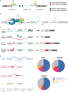
Figure 1.. Fundamentals of RNA splicing and how mutations within genes alter splicing in cis.
(A) Diagram of an intron and two flanking exons with consensus sequences defining the 5’ splice site (ss), branchpoint, and 3’ ss. Colored boxes depict sequences within exons and introns that increase or decrease the likelihood of splice site recognition by RNA binding proteins (splicing enhancers or repressors, respectively). (B) Diagram of the SF3b complex of the spliceosome (which contains SF3B1), associated RNA binding proteins (the U2AF heterodimer and an accessory splicing factor RBM39) and the sequential reactions involved in removal of an intron (intron shown in teal and exons in grey). As shown, the SF3b complex is involved in recognizing the branchpoint (shown here as an adenosine nucleotide (“A”)) and is recruited to this site by the U2AF complex, which recognizes sequences at the 3’ ss. During splicing catalysis, the branchpoint A carries out a nucleophilic attack at the 5’ ss, forming a lariat, and then the 3’OH of the released 5’ exon performs a second nucleophilic attack at the last nucleotide of the intron at the 3’ ss, joining the exons and releasing the intron lariat. (C) Diagram of how single nucleotide variants near splice sites, throughout an exon, and deep within introns may disrupt splicing or generate novel aberrant splice sites in the mRNA of a gene in cis. (D) Pie charts depicting distribution of each category of splicing event shown on the left based on annotations of the human genome from RefSeq, GenCode, and EnsEMBL.(130) “Other” represents complex splicing events (>1 of the five categories found simultaneously) as well as the small proportion of splicing events represented by mutually exclusive exons.
Figure 2.
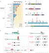
Figure 2.. Frequency, location, and global impact on RNA splicing of recurrently mutated splicing factors in cancer.
(A) Histogram depicting frequency of mutations in SF3B1, SRSF2, U2AF, and ZRSR2 in hematopoietic malignancies and solid tumors. In addition to these four genes (which are the most frequently mutated in hematopoietic malignancies), a host of additional splicing factors affected by hotspot as well as presumed loss-of-function mutations are also mutated in cancer and not shown here. Abbreviations: RARS (refractory anemia with ring sideroblasts), RCMD-RS (refractory cytopenias with multilineage dysplasia and ring sideroblasts), MDS (myelodysplastic syndromes), CMML (chronic myelomonocytic leukemia), AML-MRC (acute myeloid leukemia with myelodysplasia related changes), CLL (chronic lymphocytic leukemia), LUAD (lung adenocarcinoma). (B) Protein diagrams of the four mutated splicing factors shown in (A) with location of mutant residues. Hotspot mutations are shown in red. Abbreviations: HD (HEAT repeat domain), Zn (Zinc finger), RRM (RNA recognition motif), RS (Serine/Arginine rich domain), UHM (U2AF homology motif). (C) Diagram of an intron, two flanking exons, and locations in RNA where the four factors from (A) bind (top left). Mutations in SRSF2 skew the binding avidity of SRSF2 such that mutants bind C-rich sequences more avidly to promote exon splicing while reducing binding affinity for G rich sequences (middle left). SF3B1 is responsible for recognition of the branchpoint. Mutations in SF3B1 cause recognition of an aberrant branchpoint leading to intron proximal alternative 3’ ss selection (bottom left). Finally, U2AF1 is responsible for recognition of the 3’ yAG|r dinucleotide (where “y” represents the C- or T- pyrimidine nucleotide immediately intronic to the AG and r represents the first nucleotide in the downstream exon (at the +1 position)). As shown on the right, U2AF1 S34F/Y mutations favor inclusion of cassette exons bearing a 3’ ss containing a C-nucleotide at the −3 position while Q157 mutants promote splicing of exons with G nucleotides at the +1 position. Panels A and B adapted from Dvinge et al., RNA splicing factors as oncoproteins and tumour suppressors. Nat Rev Cancer. 2016 Jul;16(7):413–30 and used with permission.
Figure 3.
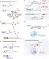
Figure 3.. Altered gene regulation in cancer through alternative cleavage and polyadenylation of mRNAs.
(A) Schematic of how alternative polyadenylation (APA) of mRNAs in the 3’-untranslated region (3’-UTR) of mRNAs results in two distinct isoforms which differ only in their 3’-UTRs. The 3’-UTR can contain multiple potential polyadenylation sites (PASs) and additional sequences that may be recognized by RNA binding proteins (RBPs) and/or micro-RNAs (miRNAs). Altering 3’-UTR length may impact miRNA-mediated gene repression, protein-protein interactions, mRNA stability, translation, export, and localization. (B) Cyclin D1 upregulation is a well-studied example of how altering 3’-UTR length results in proto-oncogene activation. In a proportion of mantle cell lymphoma patients, Cyclin D1 is upregulated through polymorphisms and mutations in the 3’-UTR that result in the use of a proximal PAS and a shortened 3’-UTR that lacks a miRNA seed region. (C) Schematic illustrating how shortening of the 3’-UTR of an mRNA may allow release of miRNAs to suppress the expression of other mRNAs in trans. (D) APA may also utilize PAS upstream of the normal stop codon and thereby alter the coding sequence of mRNA. In this example, a proximal PAS site is located within an intron and use of this PAS site results in production of a shorter protein with a novel 3’ amino acid sequence. (E) In addition to alterations in 3’-UTR length, cyclin D1 is also subject to use of upstream PASs. Polymorphisms at the end of exon 4 (for example, the G870A polymorphism) may promote use of an intronic polyadenylation signal within intron 4. This cleaves both miRNA binding sites as well as sequences encoding the normal nuclear export signal (NES) from cyclin D1. As a result, this cyclin D1 protein isoform is restricted to the nucleus.
Figure 4.
Figure 4.. Methods for therapeutic modulation of RNA splicing.
Pharmacologic means to perturb splicing include (A) drugs that physically bind the SF3b complex and disrupt its ability to recognize the branchpoint region of the intron. (B) More recently, anticancer sulfonamide compounds were discovered to cause the degradation of RBM39. These compounds physically link RBM39 to the DCAF15-CUL4 ubiquitin ligase, resulting in ubiquitinylation of RBM39 and its subsequent proteasomal degradation (of note, it is currently unknown whether degradation of RBM39 occurs while bound to U2AF and/or assembled on a 3’ splice site region). Specific mutations in SF3B1, PHF5A, and RBM39 that confer drug resistance to these molecules are shown. (C) The function, cellular localization, and assembly of a variety of splicing proteins depend on post-translational modifications and inhibitors of the enzymes placing these marks have been developed. These include protein arginine methyltransferase (PRMT) inhibitors as well as inhibitors of CLK, SRPK, and DYRK kinases. (D) Finally, oligonucleotides that modify splicing of specific transcripts by blocking the RNA–RNA base-pairing or protein–RNA binding interactions that occur between the splicing machinery and the pre-mRNA may be used to target individual aberrant splicing events in cancer.

References

    1. Han L, Diao L, Yu S, Xu X, Li J, Zhang R, et al. The Genomic Landscape and Clinical Relevance of A-to-I RNA Editing in Human Cancers. Cancer cell 2015;28:515–28. - PMC - PubMed
    1. Schmitt AM, Chang HY. Long Noncoding RNAs in Cancer Pathways. Cancer cell 2016;29:452–63. - PMC - PubMed
    1. Vu LP, Cheng Y, Kharas MG. The Biology of m(6)A RNA Methylation in Normal and Malignant Hematopoiesis. Cancer discovery 2019;9:25–33. - PubMed
    1. Peng Y, Croce CM. The role of MicroRNAs in human cancer. Signal Transduct Target Ther 2016;1:15004. - PMC - PubMed
    1. Lee Y, Rio DC. Mechanisms and Regulation of Alternative Pre-mRNA Splicing. Annu Rev Biochem 2015;84:291–323. - PMC - PubMed

Publication types

MeSH terms