

Staphylococcus aureus exhibits heterogeneous siderophore production within the vertebrate host
- PMID: 31611408
- PMCID: PMC6825271
- DOI: 10.1073/pnas.1913991116
Staphylococcus aureus exhibits heterogeneous siderophore production within the vertebrate host
Abstract
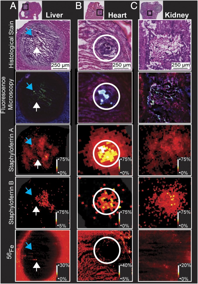
Siderophores, iron-scavenging small molecules, are fundamental to bacterial nutrient metal acquisition and enable pathogens to overcome challenges imposed by nutritional immunity. Multimodal imaging mass spectrometry allows visualization of host-pathogen iron competition, by mapping siderophores within infected tissue. We have observed heterogeneous distributions of Staphylococcus aureus siderophores across infectious foci, challenging the paradigm that the vertebrate host is a uniformly iron-depleted environment to invading microbes.
Keywords: infectious disease; metallophore; mulitmodal molecular imaging; nutritional immunity; siderophore.
Copyright © 2019 the Author(s). Published by PNAS.
Conflict of interest statement
The authors declare no competing interest.
Figures

References
-
- Andreini C., Bertini I., Cavallaro G., Holliday G. L., Thornton J. M., Metal ions in biological catalysis: From enzyme databases to general principles. J. Biol. Inorg. Chem. 13, 1205–1218 (2008). - PubMed
-
- Weinberg E. D., Iron and susceptibility to infectious disease. Science 184, 952–956 (1974). - PubMed
-
- Aisen P., Leibman A., Zweier J., Stoichiometric and site characteristics of the binding of iron to human transferrin. J. Biol. Chem. 253, 1930–1937 (1978). - PubMed
-
- Neilands J. B., A crystalline organo-iron pigment from a rust fungus (Ustilago sphaerogena). J. Am. Chem. Soc. 74, 4846–4847 (1952).
-
- Sheldon J. R., Heinrichs D. E., Recent developments in understanding the iron acquisition strategies of gram positive pathogens. FEMS Microbiol. Rev. 39, 592–630 (2015). - PubMed
Publication types
MeSH terms
Substances
Associated data
Grants and funding
LinkOut - more resources
Full Text Sources
Other Literature Sources
Molecular Biology Databases
