

Intermediate filaments and IF-associated proteins: from cell architecture to cell proliferation
- PMID: 31611503
- PMCID: PMC6819152
- DOI: 10.2183/pjab.95.034
Intermediate filaments and IF-associated proteins: from cell architecture to cell proliferation
Abstract
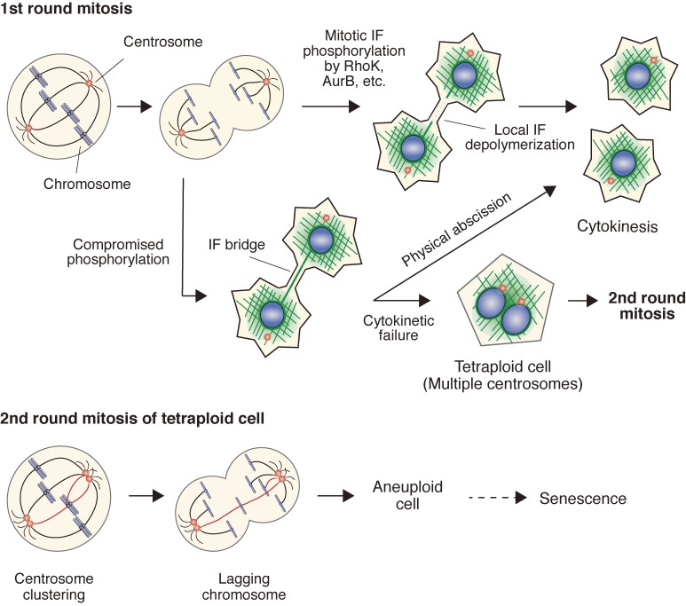
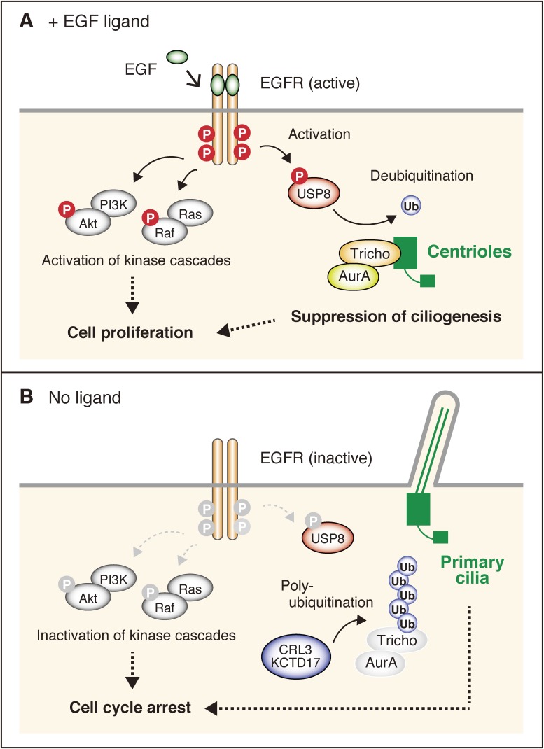
Intermediate filaments (IFs), in coordination with microfilaments and microtubules, form the structural framework of the cytoskeleton and nucleus, thereby providing mechanical support against cellular stresses and anchoring intracellular organelles in place. The assembly and disassembly of IFs are mainly regulated by the phosphorylation of IF proteins. These phosphorylation states can be tracked using antibodies raised against phosphopeptides in the target proteins. IFs exert their functions through interactions with not only structural proteins, but also non-structural proteins involved in cell signaling, such as stress responses, apoptosis, and cell proliferation. This review highlights findings related to how IFs regulate cell division through phosphorylation cascades and how trichoplein, a centriolar protein originally identified as a keratin-associated protein, regulates the cell cycle through primary cilium formation.
Keywords: cell proliferation; cytokinesis; intermediate filament; post-translational modification; primary cilium; site- and phosphorylation state-specific antibody.
Figures
References
-
- Inagaki M., Nishi Y., Nishizawa K., Matsuyama M., Sato C. (1987) Site-specific phosphorylation induces disassembly of vimentin filaments in vitro. Nature 328, 649–652. - PubMed
-
- Inagaki N., Ito M., Nakano T., Inagaki M. (1994) Spatiotemporal distribution of protein kinase and phosphatase activities. Trends Biochem. Sci. 19, 448–452. - PubMed
-
- Inagaki M., Matsuoka Y., Tsujimura K., Ando S., Tokui T., Takahashi T., et al. (1996) Dynamic property of intermediate filaments: Regulation by phosphorylation. BioEssays 18, 481–487.
Publication types
MeSH terms
Substances
LinkOut - more resources
Full Text Sources
Miscellaneous