. 2019 Oct 14;9(1):14747.
doi: 10.1038/s41598-019-50619-w.

Molecular insights into the interaction of hemorphin and its targets

Affiliations

Molecular insights into the interaction of hemorphin and its targets

Amanat Ali et al. Sci Rep. .

Abstract

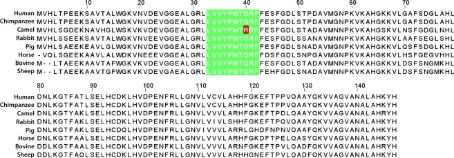
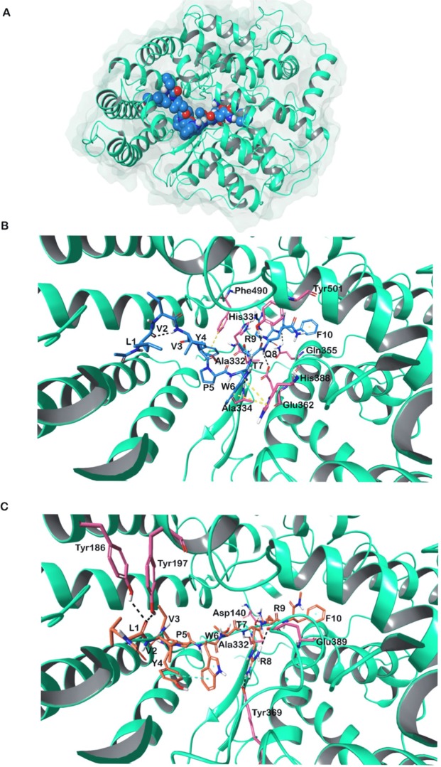
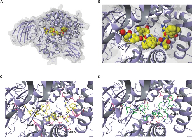
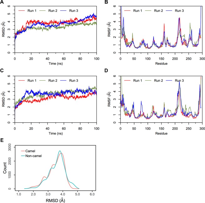
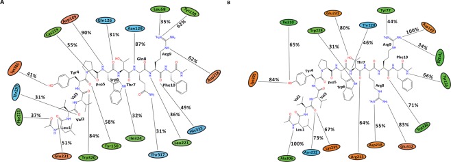
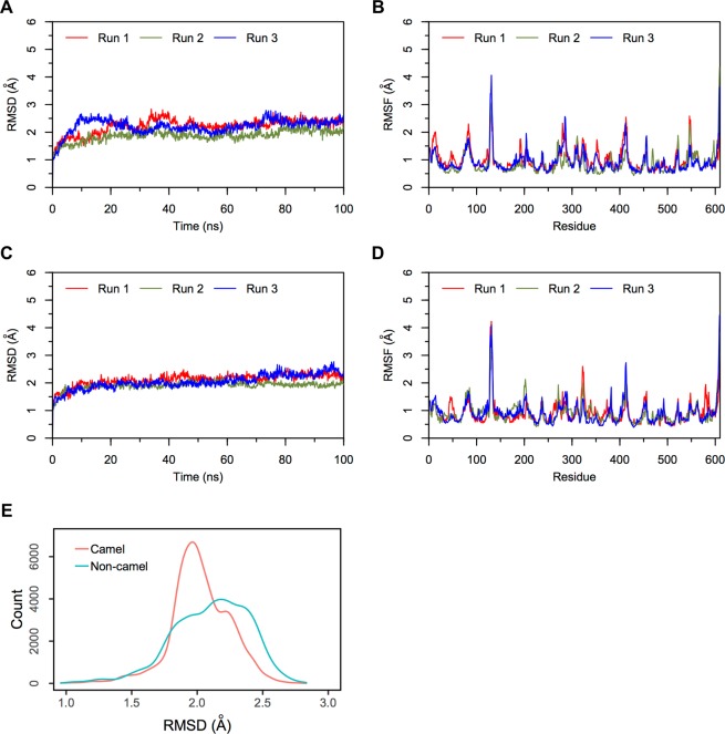
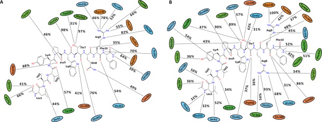
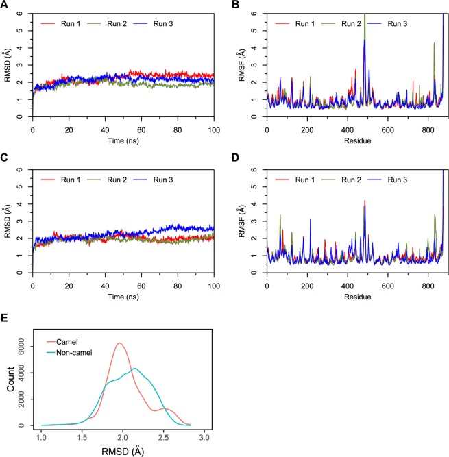
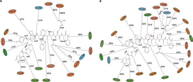
Hemorphins are atypical endogenous opioid peptides produced by the cleavage of hemoglobin beta chain. Several studies have reported the therapeutic potential of hemorphin in memory enhancement, blood regulation, and analgesia. However, the mode of interaction of hemorphin with its target remains largely elusive. The decapeptide LVV-hemorphin-7 is the most stable form of hemorphin. It binds with high affinity to mu-opioid receptors (MOR), angiotensin-converting enzyme (ACE) and insulin-regulated aminopeptidase (IRAP). In this study, computational methods were used extensively to elucidate the most likely binding pose of mammalian LVV-hemorphin-7 with the aforementioned proteins and to calculate the binding affinity. Additionally, alignment of mammalian hemorphin sequences showed that the hemorphin sequence of the camel harbors a variation - a Q > R substitution at position 8. This study also investigated the binding affinity and the interaction mechanism of camel LVV-hemorphin-7 with these proteins. To gain a better understanding of the dynamics of the molecular interactions between the selected targets and hemorphin peptides, 100 ns molecular dynamics simulations of the best-ranked poses were performed. Simulations highlighted major interactions between the peptides and key residues in the binding site of the proteins. Interestingly, camel hemorphin had a higher binding affinity and showed more interactions with all three proteins when compared to the canonical mammalian LVV-hemorphin-7. Thus, camel LVV-hemorphin-7 could be explored as a potent therapeutic agent for memory loss, hypertension, and analgesia.


Conflict of interest statement

The authors declare no competing interests.

Figures

Figure 1
Figure 1
Multiple sequence alignment of hemoglobin beta protein sequences from closely related mammals - Homo sapiens (human), Pan troglodytes (chimpanzee), Camelus dromedarius (camel), Oryctolagus cuniculus (rabbit), Sus scrofa (wild pig), Equus caballus (horse), Bos taurus (bovine), and Ovis aries (sheep).
Figure 2
Figure 2
Modeled active human MOR. (A) Side view (B) Top view. (C) LVVYPWTQRF (non-camel LVV-hemorphin-7) docked in the binding pocket of the putative active conformation of human MOR. (D) LVVYPWTRRF (camel LVV-hemorphin-7) docked in the binding pocket of the putative active conformation of human MOR. Hydrogen bonds are represented by black dotted lines and π-π stacking by yellow dotted lines.
Figure 3
Figure 3
(A) Three dimensional structure of ACE. (B) LVVYPWTQRF (non-camel LVV-hemorphin-7) docked in the active site of ACE. (C) LVVYPWTRRF (camel LVV-hemorphin-7) docked in the active site of ACE. Hydrogen bonds are represented by black dotted lines and π-π stacking represented by yellow dotted lines.
Figure 4
Figure 4
(A) Three dimensional structure of IRAP. (B) Binding pocket of IRAP. (C) LVVYPWTQRF (non-camel hemorphin) docked in the binding pocket of IRAP. (D) LVVYPWTRRF (camel hemorphin) docked in the binding pocket of IRAP. Hydrogen bonds are represented by black dotted lines and cation-π interactions are represented by red dotted lines.
Figure 5
Figure 5
RMSD and RMSF plots of triplicate 100 ns simulations of MOR. Data from the three runs are plotted with red, blue and green lines. (A) RMSD of protein Cα atoms from the MOR-LVVYPWTQRF simulations. (B) RMSF of protein Cα atoms from the MOR-LVVYPWTQRF simulations. (C) RMSD of protein Cα atoms from the MOR-LVVYPWTRRF simulations. (D) RMSF of protein Cα atoms from the MOR-LVVYPWTRRF simulations. (E) Density functions corresponding to the distribution of RMSD values from triplicate hemorphin-bound simulations.
Figure 6
Figure 6
Average percentage of equilibrium simulation time during which MOR residues maintain contact with non-camel and camel LVV-hemorphin-7 from three 100 ns simulations. For equilibrium simulation data, the first 50 ns of run 1 was discarded, while the first 30 ns of runs 2 and 3 were discarded. Histograms representing the interaction from each of the 3 simulations can be found in Supplementary Fig. 5. Charged, hydrophobic and polar amino acids are represented with orange, green and blue color respectively. (A) Average percentage of time an MOR residue maintains contact with LVVYPWTQRF. (B) Average percentage of time an MOR residue maintains contact with LVVYPWTRRF.
Figure 7
Figure 7
RMSD and RMSF plots of triplicate 100 ns simulations of ACE. Data from the three runs are plotted with red, blue and green lines. (A) RMSD of protein Cα atoms from the ACE-LVVYPWTQRF simulations. (B) RMSF of protein Cα atoms from the ACE-LVVYPWTQRF simulations. (C) RMSD of protein Cα atoms from the ACE-LVVYPWTRRF simulations. (D) RMSF of protein Cα atoms from the ACE-LVVYPWTRRF simulations. (E) Density functions corresponding to the distribution of RMSD values from triplicate hemorphin-bound simulations.
Figure 8
Figure 8
Average percentage of equilibrium simulation time during which ACE residues maintain contact with non-camel and camel LVV-hemorphin-7 from three 100 ns simulations. For equilibrium simulation data, the first 30 ns of each of the three simulations were discarded. Histograms representing the interaction from each of the 3 simulations can be found in Supplementary Fig. 6. Charged, hydrophobic and polar amino acids are represented with orange, green and blue color respectively. (A) Average percentage of time an ACE residue maintains contact with LVVYPWTQRF. (B) Average percentage of time an ACE residue maintains contact with LVVYPWTRRF.
Figure 9
Figure 9
RMSD and RMSF plots of triplicate 100 ns simulations of IRAP. Data from the three runs are plotted with red, blue and green lines. (A) RMSD of protein Cα atoms from the IRAP-LVVYPWTQRF simulations. (B) RMSF of protein Cα atoms from the IRAP-LVVYPWTQRF simulations. (C) RMSD of protein Cα atoms from the IRAP-LVVYPWTRRF simulations. (D) RMSF of protein Cα atoms from the IRAP-LVVYPWTRRF simulations. (E) Density functions corresponding to the distribution of RMSD values from triplicate hemorphin-bound simulations.
Figure 10
Figure 10
Average percentage of equilibrium simulation time during which IRAP residues maintain contact with non-camel and camel LVV-hemorphin-7 from three 100 ns simulations. For equilibrium simulation data, the first 30 ns of each of the three simulations were discarded. Histograms representing the interaction from each of the 3 simulations can be found in Supplementary Fig. 7. Charged, hydrophobic and polar amino acids are represented with orange, green and blue color respectively. (A) Average percentage of time an IRAP residue maintains contact with LVVYPWTQRF. (B) Average percentage of time an IRAP residue maintains contact with LVVYPWTRRF.

References

    1. Brantl V, et al. Novel opioid peptides derived from hemoglobin: hemorphins. Eur. J. Pharmacol. 1986;125:309–310. doi: 10.1016/0014-2999(86)90044-0. - DOI - PubMed
    1. Ivanov VT, Karelin AA, Philippova MM, Nazimov IV, Pletnev VZ. Hemoglobin as a source of endogenous bioactive peptides: The concept of tissue‐specific peptide pool. Pept. Sci. 1997;43:171–188. doi: 10.1002/(SICI)1097-0282(1997)43:2<171::AID-BIP10>3.0.CO;2-O. - DOI - PubMed
    1. Nyberg F, Sanderson K, Glamsta EL. The hemorphins: a new class of opioid peptides derived from the blood protein hemoglobin. Biopolymers. 1997;43:147–156. doi: 10.1002/(SICI)1097-0282(1997)43:2<147::AID-BIP8>3.0.CO;2-V. - DOI - PubMed
    1. Fruitier, I., Garreau, I. & Piot, J.-M. Cathepsin D Is a Good Candidate for the Specific Release of a Stable Hemorphin from Hemoglobin In Vivo: VV-Hemorphin-7. Biochem. Biophys. Res. Commun. 246, 719–724 (1998). - PubMed
    1. Glamsta EL, Meyerson B, Silberring J, Terenius L, Nyberg F. Isolation of a hemoglobin-derived opioid peptide from cerebrospinal fluid of patients with cerebrovascular bleedings. Biochem. Biophys. Res. Commun. 1992;184:1060–1066. doi: 10.1016/0006-291X(92)90699-L. - DOI - PubMed

Publication types