. 2019 Oct 14;9(1):14754.
doi: 10.1038/s41598-019-51343-1.

Hypoxia-inducible factor-1α is the therapeutic target of the SGLT2 inhibitor for diabetic nephropathy

Affiliations

Hypoxia-inducible factor-1α is the therapeutic target of the SGLT2 inhibitor for diabetic nephropathy

Ryoichi Bessho et al. Sci Rep. .

Abstract

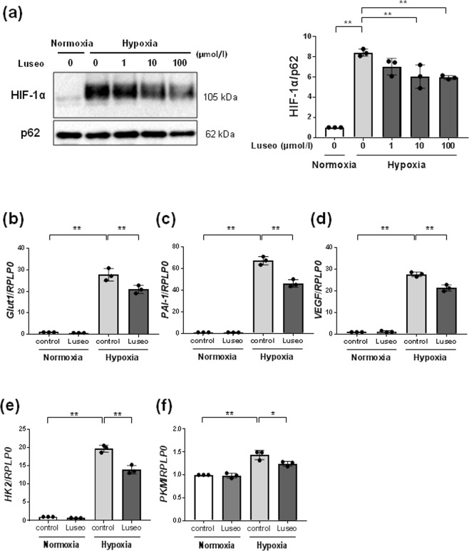
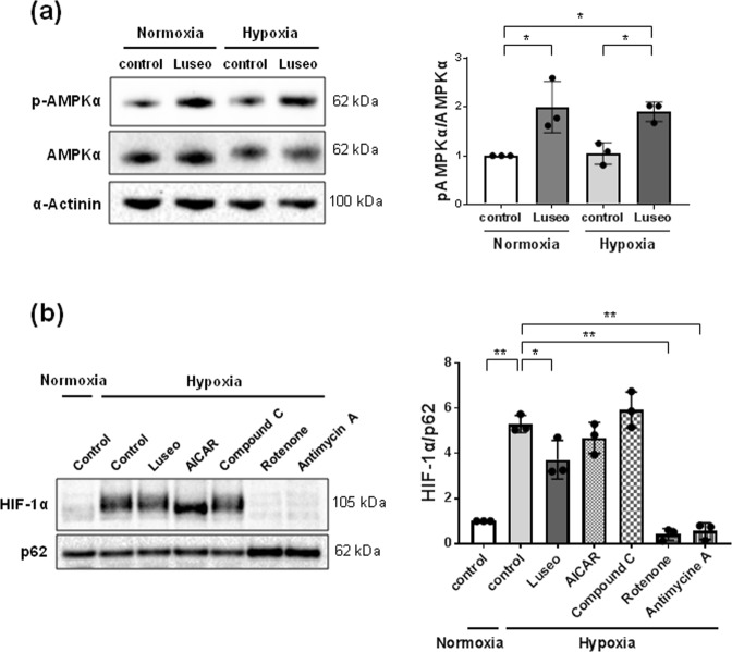
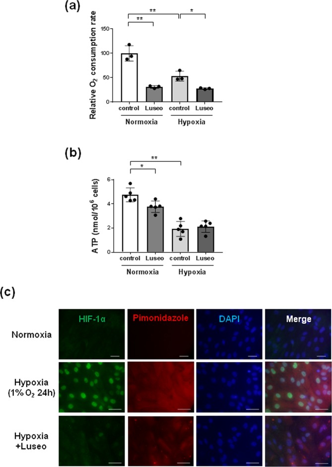
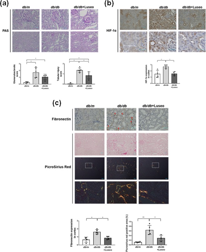
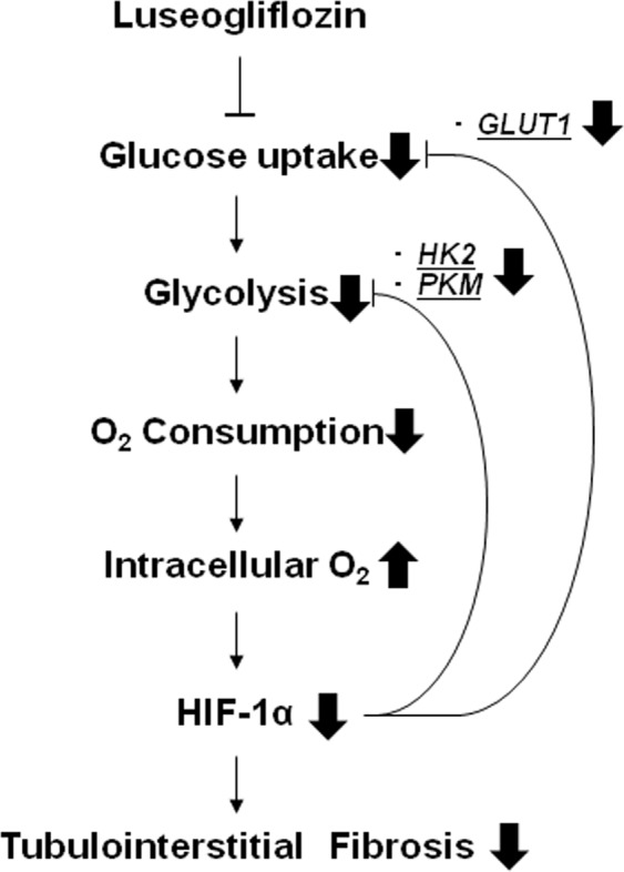
Previous studies have demonstrated intrarenal hypoxia in patients with diabetes. Hypoxia-inducible factor (HIF)-1 plays an important role in hypoxia-induced tubulointerstitial fibrosis. Recent clinical trials have confirmed the renoprotective action of SGLT2 inhibitors in diabetic nephropathy. We explored the effects of an SGLT2 inhibitor, luseogliflozin on HIF-1α expression in human renal proximal tubular epithelial cells (HRPTECs). Luseogliflozin significantly inhibited hypoxia-induced HIF-1α protein expression in HRPTECs. In addition, luseogliflozin inhibited hypoxia-induced the expression of the HIF-1α target genes PAI-1, VEGF, GLUT1, HK2 and PKM. Although luseogliflozin increased phosphorylated-AMP-activated protein kinase α (p-AMPKα) levels, the AMPK activator AICAR did not changed hypoxia-induced HIF-1α expression. Luseogliflozin suppressed the oxygen consumption rate in HRPTECs, and subsequently decreased hypoxia-sensitive dye, pimonidazole staining under hypoxia, suggesting that luseogliflozin promoted the degradation of HIF-1α protein by redistribution of intracellular oxygen. To confirm the inhibitory effect of luseogliflozin on hypoxia-induced HIF-1α protein in vivo, we treated male diabetic db/db mice with luseogliflozin for 8 to 16 weeks. Luseogliflozin attenuated cortical tubular HIF-1α expression, tubular injury and interstitial fibronectin in db/db mice. Together, luseogliflozin inhibits hypoxia-induced HIF-1α accumulation by suppressing mitochondrial oxygen consumption. The SGLT2 inhibitors may protect diabetic kidneys by therapeutically targeting HIF-1α protein.


Conflict of interest statement

The authors declare no competing interests.

Figures

Figure 1
Figure 1
Effects of luseogliflozin on hypoxia-induced HIF-1α protein and HIF-1α target gene expression. (a) Luseogliflozin inhibits hypoxia-induced HIF-1α protein expression. HRPTECs were incubated in serum-free DMEM with 1–100 µmol/l luseogliflozin under normoxic (21% O2) or hypoxic (1% O2) conditions for 24 h. The protein expression of HIF-1α was determined by western blot analysis and quantified by densitometry, with p62 as the loading control (n = 3). All protein levels are expressed as fold of control. (bf) Quantitative real-time RT-PCR analysis of HIF-1 target genes. HRPTECs were treated with or without 100 µmol/l luseogliflozin under normoxic and hypoxic conditions for 24 h. Total RNA was extracted from HRPTECs and used for quantitative RT-PCR (n = 3). The relative amounts of GLUT 1, PAI-1, VEGF, HEK2 and PKM mRNA were normalized to RPLP0 and expressed as an arbitrary unit in which the control group value equaled 1. All results are shown as the means ± SD. *p < 0.05, **p < 0.01, by one-way ANOVA followed by Tukey’s multiple comparison test.
Figure 2
Figure 2
Luseogliflozin inhibits the hypoxia-induced HIF-1α protein, independent of AMPK activation. (a) Protein levels of pAMPKα were determined by western blot analysis and quantitated by densitometry (n = 3). HRPTECs were treated with 100 μmol/l luseogliflozin under normoxic or hypoxic conditions for 24 h. Then, total cellular extracts from HRPTECs were analyzed by western blot analysis and quantified by densitometry, with α-actinin as the loading control (n = 3). Luseogliflozin promoted the phosphorylation of AMPK under normoxia and hypoxia. (b) The inhibitors of mitochondrial respiratory complexes I and III, but not the AMPK activator and inhibitor, inhibited hypoxia-induced HIF-1α accumulation in HRPTECs. HRPTECs were treated with AICAR (1 mmol/l), compound C (20 µmol/l), rotenone (1 μmol/l) and antimycin A (10 ng/mL) under hypoxic conditions for 24 h. Nuclear extracts from HRPTECs were analyzed by western blot analysis and quantified by densitometry, with p62 as the loading control (n = 3). All results are shown as the means ± SD. *p < 0.05, **p < 0.01, by one-way ANOVA followed by Tukey’s multiple comparison test.
Figure 3
Figure 3
Luseogliflozin suppressed oxygen consumption and restored intracellular hypoxia in HRPTECs. (a) The oxygen consumption rate (OCR) of HRPTECs was measured as described in the methods. Luseogliflozin (100 µmol/l) inhibited the OCR in HRPTECs under normoxic conditions. Hypoxia significantly decreased the OCR, and luseogliflozin decreased the OCR, even under hypoxic conditions. All OCR levels are expressed as fold of control (n = 3). (b) Cell ATP levels during luseogliflozin treatment under normoxia and hypoxia. HRPTECs were treated with luseogliflozin for 24 h. At the end of the incubation, cells were extracted with perchloric acid for the measurement of ATP as described in the methods (n = 5). Hypoxia significantly decreased intracellular ATP, and luseogliflozin failed to decrease ATP under hypoxic conditions. All results are shown as the means ± SD. *p < 0.05, **p < 0.01, by one-way ANOVA followed by Tukey’s multiple comparison test. (c) Immunofluorescence analysis of HIF-1α and pimonidazole in HRPTECs. HRPTECs were grown on coverslides and then treated for 24 h. Hypoxia induced the nuclear expression of HIF-1α in HRPTECs, and luseogliflozin (100 µmol/l) inhibited hypoxia-induced HIF-1α expression. Hypoxia in HRPTECs was detected by pimonidazole hydrochloride. Luseogliflozin increased cellular oxygen levels in HRPTECs under hypoxic conditions. The nuclei were stained with DAPI. Scale bars, 30 µm for normoxia and 43.1 µm for hypoxia.
Figure 4
Figure 4
Luseogliflozin ameliorates tubular injury in db/db mice, accompanied by the inhibition of HIF-1α and tubulointerstitial fibrosis. (a) Periodic acid-Schiff (PAS) staining of the glomerular tuft area surrounded by the proximal tubules in each group of mice. Scale bars, 30 µm. (b) Immunohistochemistry for HIF-1 α protein. Scale bars, 30 µm. (c) Immunohistochemistry for fibronectin and Picrosirius Red staining. The red arrows show immunoreactive staining for fibronectin in db/db mice. Scale bars, 30 μm in the top and the second panels, and 50 μm in the middle panels. Bottom panels show higher magnification images of Picrosirius Red staining in the middle panels under polarized light. Data are semiquantitative morphometric analyses of the glomerulosclerotic score and tubular injury score (a), HIF-1α (b) and fibronectin expression (c). Comparisons by Kruskal-Wallis test followed by Man-Whitney U test for multiple comparisons. Picrosirius Red staining (c) was analyzed by one-way ANOVA, Tukey’s post hoc test. *p < 0.05, **p < 0.01. Db/m mice (n = 4), db/db mice (n = 5) and luseogliflozin-treated db/db mice (n = 4).
Figure 5
Figure 5
The renoprotective mechanism of the SGLT2 inhibitor occurs through oxygen metabolism in diabetic kidneys. Because luseogliflozin inhibits glucose uptake, which leads to subsequent glycolysis and mitochondrial respiration, luseogliflozin decreases oxygen consumption in renal proximal tubular cells. Subsequently, luseogliflozin-induced intracellular oxygen redistribution supplies oxygen for prolyl hydroxylase, which promotes HIF-1α degradation in the proteasome. Consequently, luseogliflozin inhibits hypoxia-induced HIF-1α protein expression and HIF-1-induced renal fibrosis in diabetic kidneys.

References

    1. Gilbertson DT, et al. Projecting the number of patients with end-stage renal disease in the United States to the year 2015. J Am Soc Nephrol. 2005;16:3736–3741. doi: 10.1681/ASN.2005010112. - DOI - PubMed
    1. Wanner C, et al. Empagliflozin and Progression of Kidney Disease in Type 2 Diabetes. N Engl J Med. 2016;375:323–334. doi: 10.1056/NEJMoa1515920. - DOI - PubMed
    1. Neal B, et al. Canagliflozin and Cardiovascular and Renal Events in Type 2 Diabetes. N Engl J Med. 2017;377:644–657. doi: 10.1056/NEJMoa1611925. - DOI - PubMed
    1. Wiviott SD, et al. Dapagliflozin and Cardiovascular Outcomes in Type 2 Diabetes. N Engl J Med. 2019;380:347–357. doi: 10.1056/NEJMoa1812389. - DOI - PubMed
    1. Perkovic V, et al. Canagliflozin and Renal Outcomes in Type 2 Diabetes and Nephropathy. N Engl J Med. 2019;380:2295–2306. doi: 10.1056/NEJMoa1811744. - DOI - PubMed

MeSH terms

Substances