. 2020 Feb;34(2):427-440.
doi: 10.1038/s41375-019-0596-4. Epub 2019 Oct 14.

Integrated nuclear proteomics and transcriptomics identifies S100A4 as a therapeutic target in acute myeloid leukemia

Affiliations

Integrated nuclear proteomics and transcriptomics identifies S100A4 as a therapeutic target in acute myeloid leukemia

Bader Alanazi et al. Leukemia. 2020 Feb.

Abstract

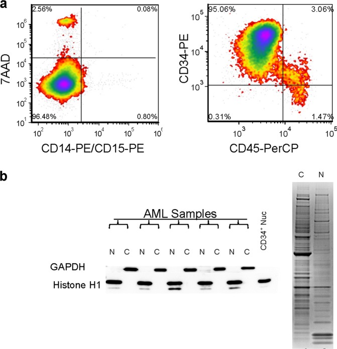
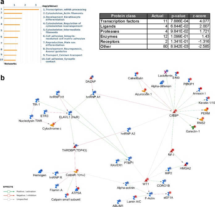
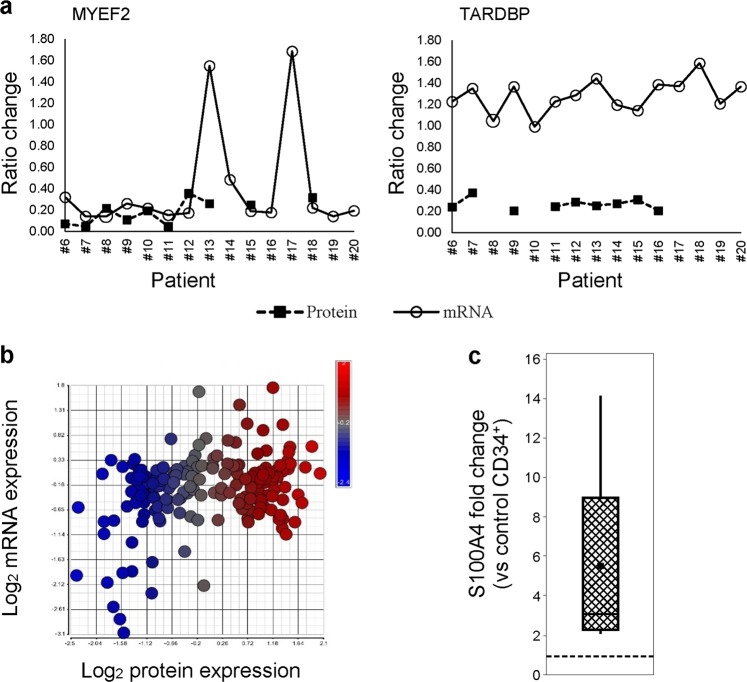
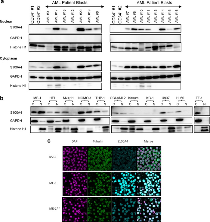
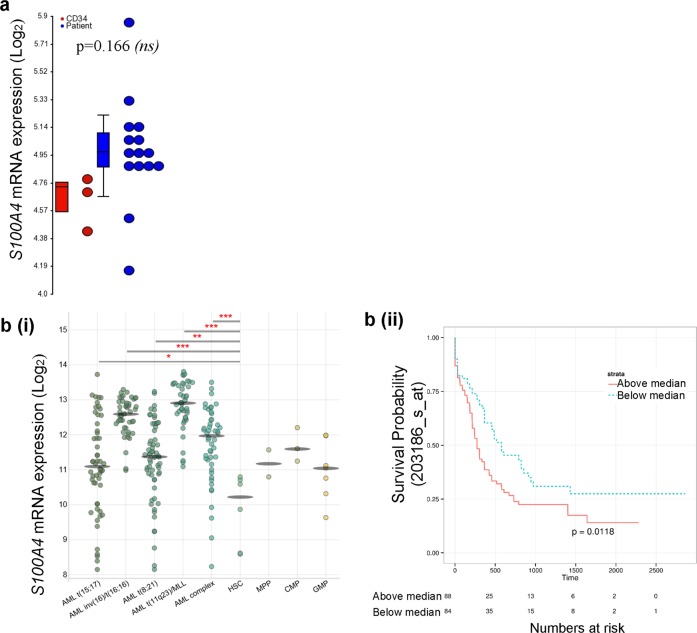
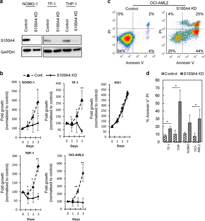
Inappropriate localization of proteins can interfere with normal cellular function and drive tumor development. To understand how this contributes to the development of acute myeloid leukemia (AML), we compared the nuclear proteome and transcriptome of AML blasts with normal human CD34+ cells. Analysis of the proteome identified networks and processes that significantly affected transcription regulation including misexpression of 11 transcription factors with seven proteins not previously implicated in AML. Transcriptome analysis identified changes in 40 transcription factors but none of these were predictive of changes at the protein level. The highest differentially expressed protein in AML nuclei compared with normal CD34+ nuclei (not previously implicated in AML) was S100A4. In an extended cohort, we found that over-expression of nuclear S100A4 was highly prevalent in AML (83%; 20/24 AML patients). Knock down of S100A4 in AML cell lines strongly impacted their survival whilst normal hemopoietic stem progenitor cells were unaffected. These data are the first analysis of the nuclear proteome in AML and have identified changes in transcription factor expression or regulation of transcription that would not have been seen at the mRNA level. These data also suggest that S100A4 is essential for AML survival and could be a therapeutic target in AML.


Conflict of interest statement

The authors declare that they have no conflict of interest.

Figures

Fig. 1
Fig. 1
Characterization and quality control of human CD34+ cells and patient AML blast samples. a Example bivariate flow cytometric plot showing viability and immunophenotype of AML blasts used in the study (left). FAB subtype (established by morphology) was confirmed by absence of CD14/CD15 expression [20]. Data exemplifying the purity of CD34+ cells is shown in the right panel. Quadrants delimit background isotype staining. b Example chromatograms of micro-capillary electrophoresis using Agilent 2100 Bioanalyzer from representative RNA samples of AML patients. c Examples of fractionated protein purity and quality. Left panel shows nitrocellulose immunoblots of samples fractioned for nuclear (N), or cytoplasmic (C) proteins. Purity of the fractioned samples was assessed by immunoblotting for GAPDH and histone protein expression. Right panel shows overall protein profile and integrity quality determined through Coomassie Brilliant Blue G staining of polyacrylamide gels
Fig. 2
Fig. 2
Functional enrichment analysis of protein changes observed in AML vs. normal hemopoietic CD34+ cells using MetacoreTM. a Enrichment analyses using Process Networks (left panel) and enrichment by protein function (right panel) shows that ‘Transcription’ is the most significant Network. The Network establishes relationships between the genes from the dataset but does not cluster them according to a specific pathway. A false discovery rate (FDR) of 0.05 was applied. b Protein networks associated with the proteins up- or downregulated in the nuclei of AML patient blasts. The network was generated using direct interaction algorithm of MetaCoreTM (Clarivate Analytics). Nodes represent proteins with lines between nodes indicating protein interactions. Only connected nodes are shown. Arrow heads indicate the direction of the interaction. Node shapes represent the functional class of the proteins as shown in the graphic key (Supplementary Fig. S2b). Red and blue circles indicate up and down regulation respectively when compared with CD34+ nuclei. Interactome analysis using “Transcription Factor” algorithm identified CEBPA (p = 5.285e−08) and WT1 (p = 0.002825) as the most significant connected transcription factors in our protein dataset
Fig. 3
Fig. 3
Correlation of protein and mRNA transcript expression of nuclear proteins changed in AML. a Protein and mRNA expression changes in two transcription factors identified to be differentially expressed between AML blasts and normal human CD34+ cells using LC/LC-MS/MS. Values below one are repressed in AML blasts. Some patients do not have a significant detection of protein when analyzed by LC/LC-MS/MS. b Correlation of nuclear protein expression with mRNA expression in nuclear proteins identified to be significantly changed between AML and CD34+ cells. Legend depicts level of change in protein expression in AML vs. normal CD34+ cells. Negative values depict lower levels of expression in AML vs. CD34+ cells. c Box and whisker plot show relative MS quantitation of S100A4 protein in expression in nuclear AML blasts vs. normal controls (n = 11). The dashed line represents no change to control (CD34+). Solid line indicates median and filled square indicates mean
Fig. 4
Fig. 4
S100A4 is over-expressed in the nucleus of AML blasts. a Example immunoblots showing validation of S100A4 protein expression and subcellular localization in same FAB M1 patient samples analyzed by MS. Supplementary Fig. S3 shows relative S100A4 expression in cytosol and nuclear fractions. S100A4 was upregulated in the nuclei and cytoplasm of 13/15 AML patients determined by western blot. AML samples 9, 11, and 12 were derived from patient bone marrow; all others AML samples were derived from peripheral blood. b Expression and subcellular localization of S100A4 in a cohort of leukemia cell lines. Cytosolic (C) and nuclear (N) fractions were analyzed by GAPDH and Histone H1 to indicate the purity/relative loading of each fraction. c Validation and expression of endogenous S100A4 expression in K562 and ME-1 leukemic cells lines using confocal laser scanning microscopy. These cell lines have either low cytoplasmic or high nuclear protein expression of S100A4 respectively. Cells were stained with DAPI and Tubulin to define cytoplasm and nuclear compartments. Fluorescence gains were equivalent (and based on isotype controls for each panel); except for ME−1** whose gain was reduced to allow the visualization of S100A4 protein expression without saturation as shown in the middle panel
Fig. 5
Fig. 5
Over-expression of S100A4 mRNA in AML. a Microarray data demonstrating the normalized intensity of S100A4 mRNA (log2) expression in normal human CD34+ cells (red; n = 3) and FAB-M1 AML (n = 15). RNA was isolated from samples that underwent mass spectrometry. The transcriptome of these samples were analyzed by Affymetrix Gene expression Profiling and data analyzed using Partek Genomics Suite v6. The Pearson correlation of S100A4 mRNA with protein expression was r = 0.45 (CI −0.206, 0.827). b S100A4 mRNA expression data from Bloodspot [69]. (b(i)) mRNA expression level of S100A4 in different AML subtypes vs. normal human hematopoietic developmental subsets. Human normal hematopoiesis data derived from GSE42519 [30] and human AML data derived from GSE13159 [70]. HSC, Hematopoietic stem cell Lin CD34+ CD38 CD90+ CD45RA; MPP, Multipotential progenitors Lin CD34+ CD38 CD90 45RA; CMP, Common myeloid progenitor cell Lin CD34+ CD38+ CD45RA- CD123+; GMP, Granulocyte monocyte progenitors Lin CD34+ CD38+ CD45RA+ CD123+. (b(ii)) Overall survival of AML patients stratified according to S100A4 expression level using the AML TCGA dataset [31]. Statistical significance is denoted by *P < 0.05; **P < 0.01 and ***P < 0.001 analyzed by t-test. ns; not significant
Fig. 6
Fig. 6
S100A4 is required for cell survival in leukemia cell lines. a Example western blot showing S100A4 expression in leukemia cells with S100A4 knocked down (KD; TRCN0000416498) compared with control (targeting nonmammalian gene) using shRNA. b Summary data showing growth of leukemia lines with S100A4 KD (TRCN0000416498) compared with control over 3 days of growth following infection (n = 3; except KG1 (n = 2). c, d Apoptosis was evaluated by flow cytometric analysis of APC-conjugated Annexin V binding, while simultaneously assessing membrane integrity by PI exclusion. c Example flow cytometric plots of S100A4 KD compared with control using OCI-AML2. Annexin V and PI negative (lower—left quadrant), annexin V+ and PI (lower—right quadrant) and both annexin V and PI positive (upper—right quadrant) cells were considered as the viable, early-phase apoptotic, late-phase apoptotic/necrotic cells, respectively. d Summary data showing the effect of S100A4 KD on Annexin V staining in leukemia cell lines following 48 h post infection. Data indicates mean ± 1 SD (n = 3). Statistical significance is denoted by *P < 0.05; **P < 0.001 analyzed by paired t-test

References

    1. Ferrara F, Schiffer CA. Acute myeloid leukaemia in adults. Lancet. 2013;381:484–95. doi: 10.1016/S0140-6736(12)61727-9. - DOI - PubMed
    1. Moarii M, Papaemmanuil E. Classification and risk assessment in AML: integrating cytogenetics and molecular profiling. Hematol Am Soc Hematol Educ Program. 2017;2017:37–44. doi: 10.1182/asheducation-2017.1.37. - DOI - PMC - PubMed
    1. Kentsis A, Reed C, Rice KL, Sanda T, Rodig SJ, Tholouli E, et al. Autocrine activation of the MET receptor tyrosine kinase in acute myeloid leukemia. Nat Med. 2012;18:1118–22. doi: 10.1038/nm.2819. - DOI - PMC - PubMed
    1. Vogel C, Marcotte EM. Insights into the regulation of protein abundance from proteomic and transcriptomic analyses. Nat Rev Genet. 2012;13:227–32. doi: 10.1038/nrg3185. - DOI - PMC - PubMed
    1. Wang X, Li S. Protein mislocalization: mechanisms, functions and clinical applications in cancer. Biochim Biophys Acta. 2014;1846:13–25. - PMC - PubMed

Publication types