. 2019 Nov;16(11):1161-1168.
doi: 10.1038/s41592-019-0591-8. Epub 2019 Oct 14.

A complete data processing workflow for cryo-ET and subtomogram averaging

Affiliations

A complete data processing workflow for cryo-ET and subtomogram averaging

Muyuan Chen et al. Nat Methods. 2019 Nov.

Abstract

Electron cryotomography is currently the only method capable of visualizing cells in three dimensions at nanometer resolutions. While modern instruments produce massive amounts of tomography data containing extremely rich structural information, data processing is very labor intensive and the results are often limited by the skills of the personnel rather than the data. We present an integrated workflow that covers the entire tomography data processing pipeline, from automated tilt series alignment to subnanometer resolution subtomogram averaging. Resolution enhancement is made possible through the use of per-particle per-tilt contrast transfer function correction and alignment. The workflow greatly reduces human bias, increases throughput and more closely approaches data-limited resolution for subtomogram averaging in both purified macromolecules and cells.


Conflict of interest statement

Competing financial interests

The authors declare no competing financial interests.

Figures

Figure 1.
Figure 1.. Diagram of CryoET data processing workflow.
(a) Main workflow diagram. (b) Workflow of tomogram reconstruction. (c) Workflow of subtomogram refinement and subtilt refinement.
Figure 2.
Figure 2.. Results of iterative tomogram alignment and reconstruction.
(a) Cellular tomogram of an E. coli bacterium with gold fiducials. (b) Selected landmark projections from (a) (left) x-y plane; (mid) x-z plane after the first iteration of the iterative alignment; (right) x-z plane after iterative alignment. (c) Tomogram of purified apoferritin without fiducials (EMPIAR-10171). (d) Selected landmark projections from (c). (e) Automatic specimen plane positioning. Left: (top) x-y slice (bottom) x-z slice, both before specimen plane positioning; right: the specimen becomes flat in the tomogram after automated positioning.
Figure 3.
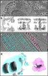
Figure 3.. Particle extraction and initial model generation.
(a) Slice view of a E. coli tomogram with particles of Tolc-AcrAB pump selected. (b) Initial model generation from Tolc-AcrAB pump particles. From the left to right are density maps of the initial seed, after 5 iterations with c1 symmetry, and after 5 iterations with c3 symmetry. (c) A tomogram slice view of the flagellum of an anucleated Trypanosoma brucei cell, with cyan circles selecting microtubule doublets, and pink circle selecting ribosomes. (d) Initial model generated from microtubule (left) doubles and ribosomes (right).
Figure 4.
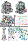
Figure 4.. Subtomogram refinement.
(a) Subtomogram averaging of ribosome (EMPIAR-10064) before subtilt refinement. (b) Subtomogram averaging after subtilt refinement. (c) Zoomed-in view of (b) with yellow arrows pointing to RNA helices and cyan arrows pointing to resolved alpha-helices. (d) Gold-standard FSC curves of the ribosome subtomogram averaging before (red) and after (blue) subtilt refinement. (e) Subtomogram averaging of the tolc-acrAB drug pump. (f) Location and orientation of the drug pump particles mapped back to a tomogram.

References

    1. Asano S, Engel BD & Baumeister W In Situ Cryo-Electron Tomography: A Post-Reductionist Approach to Structural Biology. J. Mol. Biol 428, 332–343 (2016). - PubMed
    1. Kremer JR, Mastronarde DN & McIntosh JR Computer visualization of three-dimensional image data using IMOD. J. Struct. Biol 116, 71–76 (1996). - PubMed
    1. Frangakis AS et al. Identification of macromolecular complexes in cryoelectron tomograms of phantom cells. Proc. Natl. Acad. Sci 99, 14153–14158 (2002). - PMC - PubMed
    1. Amat F et al. Markov random field based automatic image alignment for electron tomography. J. Struct. Biol 161, 260–75 (2008). - PubMed
    1. Hrabe T et al. PyTom: A python-based toolbox for localization of macromolecules in cryo-electron tomograms and subtomogram analysis. J. Struct. Biol 178, 177–188 (2012). - PubMed

Publication types

MeSH terms