. 2019 Sep 2:18:750-763.
doi: 10.17179/excli2019-1459. eCollection 2019.

Differential effects of luteolin and its glycosides on invasion and apoptosis in MDA-MB-231 triple-negative breast cancer cells

Affiliations

Differential effects of luteolin and its glycosides on invasion and apoptosis in MDA-MB-231 triple-negative breast cancer cells

Jiyon Lee et al. EXCLI J. .

Abstract

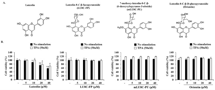
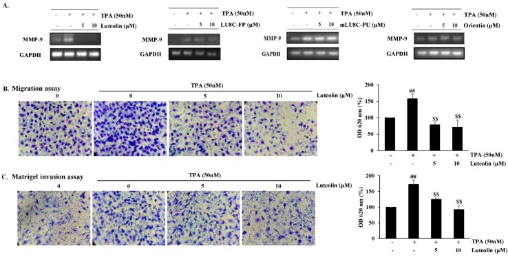
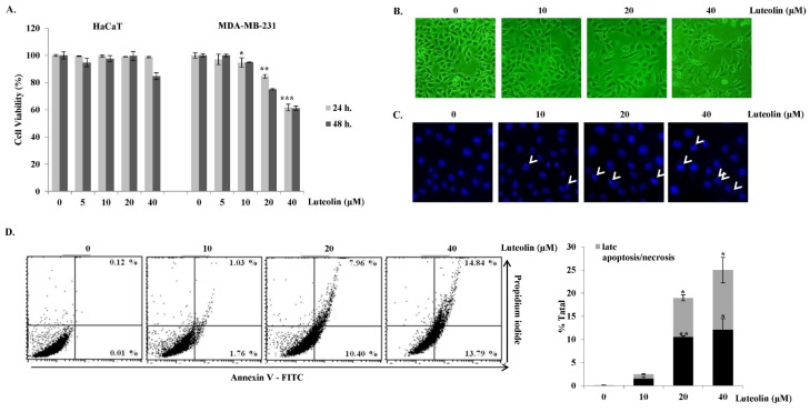
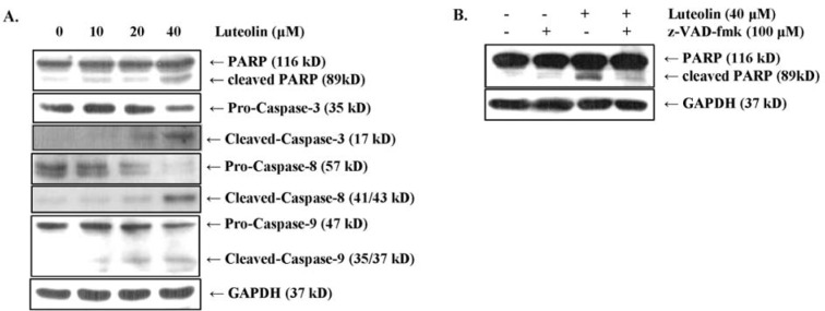
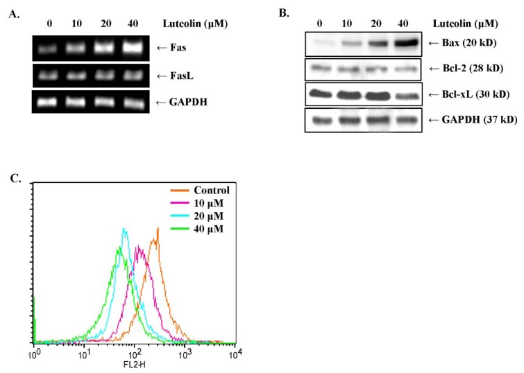
Luteolin is known to have anticancer activity in various cancers. Recent studies have shown that luteolin glycosides, such as luteolin-8-C-β-fucopyranoside, 7-methoxy-luteolin-8-C-β-(6- deoxyxylopyranos-3-uloside) and luteolin-8-C-β-d-glucopyranoside, flavonoids that are present in Arthraxon hispidus, exert antimigratory and anti-invasive effects, but no cytotoxic effect in estrogen receptor-positive MCF7 breast cancer cells. In the present study, we further investigated and compared differential effects of luteolin and its glycosides in MDA-MB-231 triple-negative breast cancer cells. Luteolin suppressed the expression of matrix metalloproteinase-9 and inhibited migration and invasion in MDA-MB-231 cells treated with the tumor promotor 12-O-tetradecanoylphorbol-13-acetate at non-cytotoxic concentrations (0, 5, and 10 μM). Furthermore, at cytotoxic concentrations (20 and 40 μM), luteolin induced apoptosis via extrinsic and intrinsic pathways in MDA-MB-231 cells. However, luteolin glycosides did not exert any cytotoxic, antimigratory, or anti-invasive effect in MDA-MB-231 cells. In brief, luteolin had both antimetastatic and cytotoxic effects on MDA-MB-231 cells, whereas luteolin glycosides had no effect on this cell line. Taking together the present results and our previous findings on the differential effects of luteolin and its glycosides on MDA-MB-231 and MCF-7 breast cancer cells, luteolin and its glycosides can be suggested as a potential candidate for breast cancer therapy.

Keywords: apoptosis; breast cancer; invasion; luteolin; tumor migration.


Figures

Figure 1
Figure 1. Effects of luteolin and its glycosides on the viability of MDA-MB-231 breast cancer cells. (A) Structures of luteolin, luteolin-8-C-β-fucopyranoside (LU8C-FP), 7-methoxy-luteolin-8-C-β-(6-deoxyxylopyranos-3-uloside) (mLU8C-PU), and luteolin-8-C-β-d-glucopyrano side (orientin). (B) MDA-MB-231 breast cancer cells were treated with luteolin or its glycosides in the presence or absence of TPA (50 nM) for 24 h. Cell viability was determined by MTS assay. Statistically significant differences between luteolin-treated versus non-treated cells were determined by two-tailed Student's t-test. *p < 0.05, **p < 0.005 (n = 3)
Figure 2
Figure 2. Luteolin inhibits migration and invasion in MDA-MB-231 breast cancer cells. MDA-MB-231 cells were pretreated with luteolin, LU8C-FP, mLU8C-PU, orientin, or TPA (50 nM) for 24 h. (A) MMP-9 mRNA expression levels were evaluated by RT-PCR. (B) Migration assay and (C) Matrigel invasion assay of TPA-stimulated MDA-MB-231 cells exposed to luteolin for 24 h. Statistical significance was analyzed by one-way ANOVA followed by Tukey's HSD test. ##p<0.005 (non-treated vs. TPA alone) and $$p < 0.005 (TPA alone vs. TPA plus luteolin) (n = 3)
Figure 3
Figure 3. Cytotoxic and apoptotic effects of luteolin in MDA-MB-231 breast cancer cells. (A) MDA-MB-231 breast cancer cells and HaCaT normal keratinocytes were treated with luteolin for 24 or 48 h. Cell viability was determined by MTS assay. (B) Morphological changes in MDA-MB-231 cells after treatment with various concentrations (0, 10, 20, and 40 μM) of luteolin for 24 h were observed under a phase-contrast microscope (100×). (C) Apoptotic nuclei after treatment with luteolin were observed under a fluorescence microscope after Hoechst staining (100×). (D) Flow-cytometric analysis after staining with Annexin V-fluorescein isothiocyanate (FITC) and propidium iodide (PI). Percentages of significant events in early apoptosis (bottom right quadrants) and late apoptosis (top right quadrants). Bar graph represents annexin V+/PI- (early apoptotic) and annexin V+/PI+ (late apoptotic) cells (n = 3). Statistically significant differences between luteolin-treated versus non-treated cells were analyzed by two-tailed Student's t-test. *p < 0.05, **p < 0.005, ***p < 0.001 (n = 3)
Figure 4
Figure 4. Luteolin induces apoptosis via the caspase cascade and PARP degradation in MDA-MB-231 breast cancer cells. (A) Processing of caspases-8, -9, and -3 and PARP were detected by Western blot analysis. (B) MDA-MB-231 breast cancer cells were pretreated with 100 μM z-VAD-fmk (pan-caspase inhibitor) for 2 h and subsequently stimulated with 40 μM luteolin. PARP levels were analyzed by Western blot analysis, and GAPDH was used as an internal control.
Figure 5
Figure 5. Luteolin induces apoptosis via the extrinsic and intrinsic pathways in MDA-MB-231 breast cancer cells. (A) mRNA expression levels of Fas death receptor and Fas ligand (FasL) were analyzed by RT-PCR. (B) Protein expression levels of Bax, Bcl-2, and Bcl-xL were detected by Western blot analysis. (C) The mitochondrial transmembrane potential (∆Ψm) was analyzed by JC-1 staining after treating the cells with luteolin for 24 h. The histogram shows the distribution of JC-1. Orange color (FL2-H, right) represents JC-1 aggregates, which are a characteristic feature of healthy cells; green color (FL2-H, left) represents JC-1 monomers, which are a characteristic feature of apoptotic cells.

References

    1. Adams JM. Ways of dying: multiple pathways to apoptosis. Genes Dev. 2003;17:2481–2495. - PubMed
    1. Ashkenazi A. Targeting the extrinsic apoptotic pathway in cancer: lessons learned and future directions. J Clin Invest. 2015;125:487–489. - PMC - PubMed
    1. Aziz N, Kim MY, Cho JY. Anti-inflammatory effects of luteolin: A review of in vitro, in vivo, and in silico studies. J Ethnopharmacol. 2018;225:342–358. - PubMed
    1. Bak Y, Ham S, Baatartsogt O, Jung SH, Choi KD, Han TY, et al. A1E inhibits proliferation and induces apoptosis in NCI-H460 lung cancer cells via extrinsic and intrinsic pathways. Mol Biol Rep. 2013;40:4507–4519. - PubMed
    1. Choi J, Gyamfi J, Jang H, Koo JS. The role of tumor-associated macrophage in breast cancer biology. Histol Histopathol. 2018;33:133–145. - PubMed

LinkOut - more resources