Review
. 2019 Sep 27:10:2166.
doi: 10.3389/fimmu.2019.02166. eCollection 2019.

Complement Inhibitors in Clinical Trials for Glomerular Diseases

Affiliations
Review

Complement Inhibitors in Clinical Trials for Glomerular Diseases

Peter F Zipfel et al. Front Immunol. .

Abstract

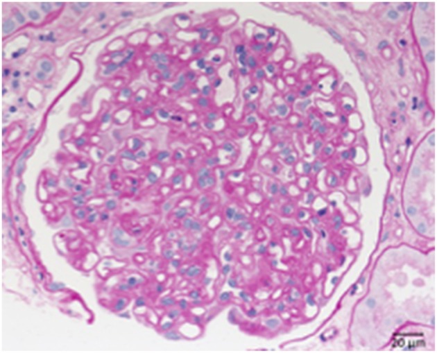
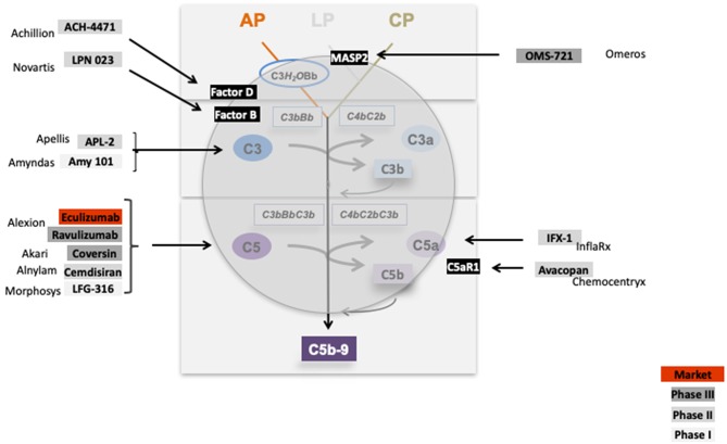
Defective complement action is a cause of several human glomerular diseases including atypical hemolytic uremic syndrome (aHUS), anti-neutrophil cytoplasmic antibody mediated vasculitis (ANCA), C3 glomerulopathy, IgA nephropathy, immune complex membranoproliferative glomerulonephritis, ischemic reperfusion injury, lupus nephritis, membranous nephropathy, and chronic transplant mediated glomerulopathy. Here we summarize ongoing clinical trials of complement inhibitors in nine glomerular diseases and show which inhibitors are used in trials for these renal disorders (http://clinicaltrials.gov).

Keywords: ANCA; C3 glomerulopathy; aHUS; clinical trials; complement; glomerular disease; inhibitors.


Figures

Figure 1
Figure 1
Overview on complement activation and cascade progression. Complement activation is mediated by the alternative pathway in the fluid phase and on surfaces, and by the lectin and the classical pathway on surfaces. Multiple regulators determine and adjust cascade progression and subsequence effector action. Thereby discriminating the action between intact self, altered self, and non-self. Three initial reactions activate the complement cascade and have different initial triggers. The alternative pathway (AP) which is initiated spontaneously and continuously in the fluid phase and in the absence of regulators is amplified on surfaces. Both the lectin (LP) and the classical pathway (CP) are initiated on surfaces. The type of surface influences activation and the regulator repertoire decides on cascade progression or inhibition. Different regulators control cascade progression in the fluid phase and on surfaces. The three pathways form surface bound convertases, the AP allows generation of the AP-C3 convertase and the LP/CP trigger CP convertase. The AP C3 convertase also triggers a potent amplification loop. The general role of both C3 convertase is to cleave the abundant plasma protein C3 (concentration 1,000–1,500 μg/ml) into the anaphylatoxin C3a and the opsonic C3b. The enzymatic response on the first enzymatic levels is frequently enhanced by the potent self-amplifying amplification loop. If activation C5 convertases are generated and again C5 convertases of the AP and of the LP/CP pathways do exist. The major role of the C5 convertase is to cleave C5 (plasma concentration 350 μg/ml) into the powerful anaphylatoxin C5a and to generate C5b. Surface bound C5b initiates terminal complement and formation of the terminal complex, C5b-9, also termed membrane attack complex which can form lytic pores. Thus, complement acts on two major enzymatic levels, each of which generates a unique set of effector components with rather diverse functions. The complexity of this cascade is mediated by regulators and inhibitors, which control activation in the fluid phase (AP) and which ensure that activation mainly occurs on non-self surfaces or modified surfaces. In the physiological setting this coordinated action allows to direct the toxic and clearance power to the foreign/modified particles. In case of any dysbalance this action can be targeted toward self-structures and this can cause pathology at specific sites.
Figure 2
Figure 2
Morphological changes in atypical uremic hemolytic syndrome (aHUS) resulting in thrombotic microangiopathy (TMA). (A) Periodic acid Schiff (PAS) reaction: focal loss of endothelial cells and fibrin precipitates in dilated glomerular capillaries (→). (B) Loss of CD34-positive endothelial cells stained red (>). (C) Accumulation of platelets stained red in CD61 immunohistochemistry (→).
Figure 3
Figure 3
Morphological changes in ANCA associated pauci-immune necrotizing glomerulonephritis. PAS-reaction of a case of ANCA-associated Glomerulonephritis, displaying a rupture of the glomerular basement membrane. See fresh fibrin precipitates inside the bowman space at the side of the necrosis (→).
Figure 4
Figure 4
Morphological appearance of C3 glomerulopathy. (A) PAS reaction reveals a membranoproliferative pattern with double contours of the GBM. (B) Strong immunohistochemical positivity for C3 at the GBM and in the mesangium. (C) Only very scant and segmental positivity for IgG.
Figure 5
Figure 5
Morphological changes in dense deposit disease (DDD). (A) PAS–reaction of a dense deposit disease case. Note the mesangial and endocapillary hypercellularity without prominent double contours of the glomerular basement membrane (→). (B) Note a strong red positivity for C3 inside the glomerular basement membrane (→). (C) The name giving electron dense deposits within the thickened glomerular basement membrane (GBM,→).
Figure 6
Figure 6
Morphological changes in IgA nephropathy: (A) IgA immunohistochemistry of a case of IgA nephropathy, displaying a noticeable mesangial positivity. (B) PAS reaction of the same glomerulus: note the mesangial matrix increase and the focal mesangial hypercellularity (arrow).
Figure 7
Figure 7
Morphological changes in immune complex membranoproliferative glomerulonephritis (MPGN). (A) PAS-reaction of a case of membranoproliferative glomerulonephritis due to a chronic hepatitis C infection. Note the lobular appearance and double contours of the GBM (→). (B) Note positivity for IgG at the side of double contours of the GBM (→).
Figure 8
Figure 8
Morphological changes in lupus nephritis, class IV. PAS reaction of a lupus nephritis class IV. Note lobular pattern with pronounced endocapillary and mesangial hypercellularity as well as thickened GBM.
Figure 9
Figure 9
Morphological changes in membranous glomeruloneprhits nephropathy. (A) PAS-reaction with slightly thickened GBM. (B) PLA2R1 immunohistochemistry of a PLA2R1-antibody associated case of membranous glomerulopathy. Note strong granular positivity for PLA2R1 at the GBM (>). (C) Electron microscopic depiction of subepithelial electron dense deposits (>).
Figure 10
Figure 10
Chronic transplant glomerulopathy. PAS: Chronic transplant glomerulopathy displaying pronounced double contours of the GBM and slight endocapillary hypercellularity.
Figure 11
Figure 11
Complement inhibitors target different levels and steps of the complement cascade. Complement inhibitors which are evaluated in clinical trials for various kidney diseases bind to different complement proteins and inhibit the cascade at different levels. C1 inhibitor binds to C1 and blocks C1 activation. OMS 721 binds to the lectin pathway protease MASP2. C3 targeting proteins include APL2 (Apellis) and AMY 101 (Amyndas). The Factor D inhibitor ACH-4471 (Achillion) binds to Factor D, a protease which cleaves in its active state Factor B. LPN023 (Novartis) a small Factor B binding protein blocks formation of the enzymatically active AP C3 Convertase. Several compounds target complement at the level of C5. Eculizumab, and the new version Ravulizumab (both Alexion) bind to C5 and block activation of the protein. Coversin is a tick derived C5 binding protein (Akari) and C5 inhibitor. C5 synthesis is blocked by Cemdisiran as an RNAi targeted strategy (Alnylam), and by LFG-316 (Novartis). The complement inflammatory C5a—C5aR1 axis is inhibited by IFX-1 (InflaRx) and Avacopan (Chemocentryx).

References

    1. Zipfel PF, Skerka C. Complement regulators and inhibitory proteins. Nat Rev Immunol. (2009) 9:729–40. 10.1038/nri2620 - DOI - PubMed
    1. Hajishengallis G, Reis ES, Mastellos DC, Ricklin D, Lambris JD. Novel mechanisms and functions of complement. Nat Immunol. (2017) 18:1288–98. 10.1038/ni.3858 - DOI - PMC - PubMed
    1. McCullough JW, Renner B, Thurman JM. The role of the complement system in acute kidney injury. Semin Nephrol. (2013) 33:543–56. 10.1016/j.semnephrol.2013.08.005 - DOI - PMC - PubMed
    1. Groopman EE, Rasouly HM, Gharavi AG. Genomic medicine for kidney disease. Nat Rev Nephrol. (2017) 14:83–104. 10.1038/nrneph.2017.167 - DOI - PMC - PubMed
    1. Bomback AS, Markowitz GS, Appel GB. Complement-mediated glomerular diseases: a tale of 3 pathways. Kidney Int Rep. (2016) 1:148–55. 10.1016/j.ekir.2016.06.005 - DOI - PMC - PubMed

Publication types

MeSH terms

Substances